Sahinis Chrysostomos, Kellis Eleftherios
Laboratory of Neuromechanics, Department of Physical Education and Sport Sciences at Serres, Aristotle University of Thessaloniki, Agios Ioannis, 62100 Serres, Greece.
J Funct Morphol Kinesiol. 2021 Sep 16;6(3):77. doi: 10.3390/jfmk6030077.
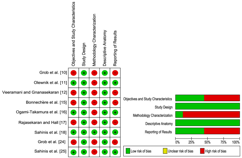
The tensor of vastus intermedius is a newly discovered muscle that is located at the anterior compartment of the thigh. The aim of the present study is to report, assess and synthetize the existing evidence on the anatomy, variation and morphological characteristics of the TVI as well as to examine its clinical importance. A systematic review was performed evaluating both anatomical and medical imaging studies which provided information about TVI anatomy, prevalence, variations and morphological characteristics. The search strategy was conducted in major electronic databases. Two reviewers worked independently to screen all possible references via a title/abstract examination. Methodological quality was examined with the Anatomical Quality Assurance checklist. A total of 295 cadaveric knees were included in the nine studies where in 244 (82.7%) cases the TVI was identified. Based on this evidence, it appears that the TVI is located between the vastus lateralis and vastus intermedius. The muscle belly is located proximally, and it is combined with a broad and flat aponeurosis before forming a tendinous structure that is attached at the medial aspect of the patella. The TVI presented some morphological variations and complex muscle architecture that varied along its length. There is insufficient good quality evidence as more than half of the included studies were ranked as having a "High" risk of bias with various methodological issues. Higher quality studies are recommended to evaluate the TVI morphology to better understand its functional and clinical importance.
股中间肌张量肌是一块新发现的肌肉,位于大腿前侧肌间隔。本研究的目的是报告、评估和综合现有的关于股中间肌张量肌(TVI)的解剖结构、变异情况和形态特征的证据,并探讨其临床重要性。进行了一项系统综述,评估了提供有关TVI解剖结构、发生率、变异情况和形态特征信息的解剖学和医学影像学研究。检索策略在主要电子数据库中进行。两名评审员独立工作,通过标题/摘要审查筛选所有可能的参考文献。采用解剖学质量保证清单检查方法学质量。九项研究共纳入295个尸体膝关节,其中244例(82.7%)发现了TVI。基于这些证据,TVI似乎位于股外侧肌和股中间肌之间。肌腹位于近端,在形成附着于髌骨内侧的腱性结构之前,它与宽阔扁平的腱膜相结合。TVI呈现出一些形态变异和复杂的肌肉结构,且沿其长度有所不同。由于超过一半的纳入研究因各种方法学问题被评为具有“高”偏倚风险,因此高质量证据不足。建议进行更高质量的研究来评估TVI的形态,以更好地理解其功能和临床重要性。