Zhang Feifei, Ran Yuncai, Zhu Ming, Lei Xiaowen, Niu Junxia, Wang Xiao, Zhang Yong, Li Shujian, Zhu Jinxia, Gao Xuemei, Mossa-Basha Mahmud, Cheng Jingliang, Zhu Chengcheng
Department of Magnetic Resonance, The First Affiliated Hospital of Zhengzhou University, Zhengzhou, China.
Department of Intervention, The First Affiliated Hospital of Zhengzhou University, Zhengzhou, China.
Front Cardiovasc Med. 2021 Sep 9;8:739332. doi: 10.3389/fcvm.2021.739332. eCollection 2021.
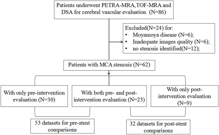
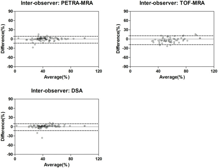
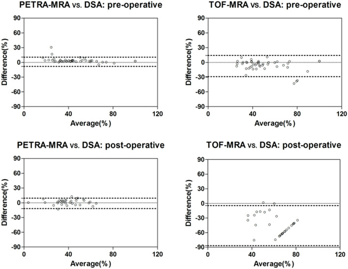
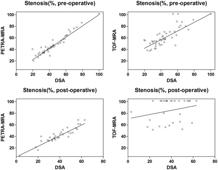
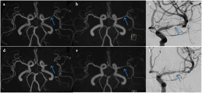
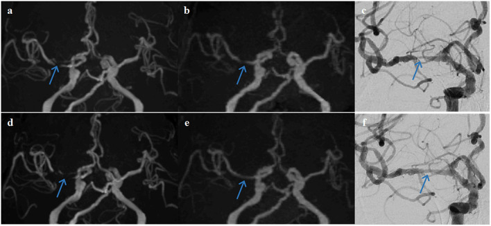
3D pointwise encoding time reduction magnetic resonance angiography (PETRA-MRA) is a promising non-contrast magnetic resonance angiography (MRA) technique for intracranial stenosis assessment but it has not been adequately validated against digital subtraction angiography (DSA) relative to 3D-time-of-flight (3D-TOF) MRA. The aim of this study was to compare PETRA-MRA and 3D-TOF-MRA using DSA as the reference standard for intracranial stenosis assessment before and after angioplasty and stenting in patients with middle cerebral artery (MCA) stenosis. Sixty-two patients with MCA stenosis (age 53 ± 12 years, 43 males) underwent MRA and DSA within a week for pre-intervention evaluation and 32 of them had intracranial angioplasty and stenting performed. The MRAs' image quality, flow visualization within the stents, and susceptibility artifact were graded on a 1-4 scale (1 = poor, 4 = excellent) independently by three radiologists. The degree of stenosis was measured by two radiologists independently on DSA and MRAs. There was an excellent inter-observer agreement for stenosis assessment on PETRA-MRA, 3D-TOF-MRA, and DSA (ICCs > 0.90). For pre-intervention evaluation, PETRA-MRA had better image quality than 3D-TOF-MRA (3.87 ± 0.34 vs. 3.38 ± 0.65, < 0.001), and PETRA-MRA had better agreement with DSA for stenosis measurements compared to 3D-TOF-MRA ( = 0.96 vs. = 0.85). For post-intervention evaluation, PETRA-MRA had better image quality than 3D-TOF-MRA for in-stent flow visualization and susceptibility artifacts (3.34 ± 0.60 vs. 1.50 ± 0.76, < 0.001; 3.31 ± 0.64 vs. 1.41 ± 0.61, < 0.001, respectively), and better agreement with DSA for stenosis measurements than 3D-TOF-MRA ( = 0.90 vs. = 0.26). 3D-TOF-MRA significantly overestimated the stenosis post-stenting compared to DSA (84.9 ± 19.7 vs. 39.3 ± 13.6%, < 0.001) while PETRA-MRA didn't (40.6 ± 13.7 vs. 39.3 ± 13.6%, = 0.18). PETRA-MRA is accurate and reproducible for quantifying MCA stenosis both pre- and post-stenting compared with DSA and performs better than 3D-TOF-MRA.
三维逐点编码时间缩短磁共振血管造影(PETRA-MRA)是一种很有前景的用于颅内狭窄评估的非对比磁共振血管造影(MRA)技术,但相对于三维时间飞跃(3D-TOF)MRARA,它尚未与数字减影血管造影(DSA)进行充分验证。本研究的目的是在大脑中动脉(MCA)狭窄患者血管成形术和支架置入术前及术后,以DSA作为颅内狭窄评估的参考标准,比较PETRA-MRA和3D-TOF-MRA。62例MCA狭窄患者(年龄53±12岁,男性43例)在一周内接受了MRA和DSA检查以进行干预前评估,其中32例患者进行了颅内血管成形术和支架置入术。三位放射科医生独立地根据1-4级标准(1 = 差,4 = 优)对MRA的图像质量、支架内血流可视化及磁敏感伪影进行分级。两位放射科医生独立地在DSA和MRA上测量狭窄程度。在PETRA-MRA、3D-TOF-MRA和DSA上狭窄评估的观察者间一致性良好(组内相关系数>0.90)。对于干预前评估,PETRA-MRA的图像质量优于3D-TOF-MRA(3.87±0.34 vs. 3.38±0.65,<0.001),并且与DSA相比PETRA-MRA在狭窄测量上与DSA的一致性优于3D-TOF-MRA(=0.96 vs. =0.85)。对于干预后评估,在支架内血流可视化和磁敏感伪影方面PETRA-MRA的图像质量优于3D-TOF-MRA(分别为3.34±0.60 vs. 1.50±0.76,<0.001;3.31±0.64 vs. 1.41±0.61,<0.001),并且在狭窄测量上与DSA的一致性优于3D-TOF-MRA(=0.90 vs. =0.26)。与DSA相比,3D-TOF-MRA在支架置入术后显著高估了狭窄程度(84.9±19.7 vs. 39.3±13.6%,<0.001),而PETRA-MRA则没有(40.6±13.7 vs. 39.3±13.6%,=0.18)。与DSA相比,PETRA-MRA在量化MCA狭窄支架置入术前及术后均准确且可重复,并且比3D-TOF-MRA表现更好。