Institute of Anatomy, University of Leipzig, 04109 Leipzig, Germany.
Department of Neurology, University of Leipzig, 04109 Leipzig, Germany.
Cells. 2021 Aug 28;10(9):2236. doi: 10.3390/cells10092236.
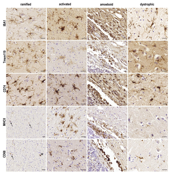
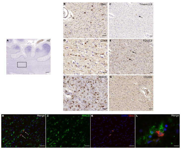
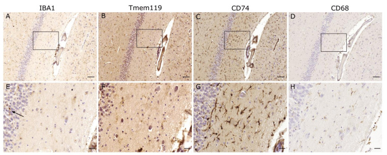
Classically, the following three morphological states of microglia have been defined: ramified, amoeboid and phagocytic. While ramified cells were long regarded as "resting", amoeboid and phagocytic microglia were viewed as "activated". In aged human brains, a fourth, morphologically novel state has been described, i.e., dystrophic microglia, which are thought to be senescent cells. Since microglia are not replenished by blood-borne mononuclear cells under physiological circumstances, they seem to have an "expiration date" limiting their capacity to phagocytose and support neurons. Identifying factors that drive microglial aging may thus be helpful to delay the onset of neurodegenerative diseases, such as Alzheimer's disease (AD). Recent progress in single-cell deep sequencing methods allowed for more refined differentiation and revealed regional-, age- and sex-dependent differences of the microglial population, and a growing number of studies demonstrate various expression profiles defining microglial subpopulations. Given the heterogeneity of pathologic states in the central nervous system, the need for accurately describing microglial morphology and expression patterns becomes increasingly important. Here, we review commonly used microglial markers and their fluctuations in expression in health and disease, with a focus on IBA1 low/negative microglia, which can be found in individuals with liver disease.
传统上,已经定义了小胶质细胞的以下三种形态状态:分支状、阿米巴样和吞噬性。虽然分支状细胞长期以来被认为是“静止的”,但阿米巴样和吞噬性小胶质细胞被视为“激活的”。在老年人大脑中,已经描述了第四种形态新颖的状态,即营养不良性小胶质细胞,它们被认为是衰老细胞。由于小胶质细胞在生理情况下不会被血液单核细胞补充,因此它们似乎有一个“到期日”,限制了它们吞噬和支持神经元的能力。因此,确定导致小胶质细胞衰老的因素可能有助于延缓阿尔茨海默病(AD)等神经退行性疾病的发作。单细胞深度测序方法的最新进展允许更精细的分化,并揭示了小胶质细胞群体的区域、年龄和性别依赖性差异,越来越多的研究表明各种定义小胶质细胞亚群的表达谱。鉴于中枢神经系统中病理状态的异质性,准确描述小胶质细胞形态和表达模式的需求变得越来越重要。在这里,我们回顾了常用的小胶质细胞标志物及其在健康和疾病中的表达波动,重点介绍了在患有肝病的个体中可以发现的 IBA1 低/阴性小胶质细胞。