• 文献检索
  • 文档翻译
  • 深度研究
  • 学术资讯
  • Suppr Zotero 插件Zotero 插件
  • 邀请有礼
  • 套餐&价格
  • 历史记录
应用&插件
Suppr Zotero 插件Zotero 插件浏览器插件Mac 客户端Windows 客户端微信小程序
定价
高级版会员购买积分包购买API积分包
服务
文献检索文档翻译深度研究API 文档MCP 服务
关于我们
关于 Suppr公司介绍联系我们用户协议隐私条款
关注我们

Suppr 超能文献

核心技术专利:CN118964589B侵权必究
粤ICP备2023148730 号-1Suppr @ 2026

文献检索

告别复杂PubMed语法,用中文像聊天一样搜索,搜遍4000万医学文献。AI智能推荐,让科研检索更轻松。

立即免费搜索

文件翻译

保留排版,准确专业,支持PDF/Word/PPT等文件格式,支持 12+语言互译。

免费翻译文档

深度研究

AI帮你快速写综述,25分钟生成高质量综述,智能提取关键信息,辅助科研写作。

立即免费体验

9.4特斯拉下基底神经节和丘脑的定量易感性图谱

Quantitative Susceptibility Mapping of the Basal Ganglia and Thalamus at 9.4 Tesla.

作者信息

Kumar Vinod Jangir, Scheffler Klaus, Hagberg Gisela E, Grodd Wolfgang

机构信息

Max Planck Institute for Biological Cybernetics, Tübingen, Germany.

Biomedical Magnetic Resonance, University Hospital and Eberhard-Karl's University, Tübingen, Germany.

出版信息

Front Neuroanat. 2021 Sep 16;15:725731. doi: 10.3389/fnana.2021.725731. eCollection 2021.

DOI:10.3389/fnana.2021.725731
PMID:34602986
原文链接:https://pmc.ncbi.nlm.nih.gov/articles/PMC8483181/
Abstract

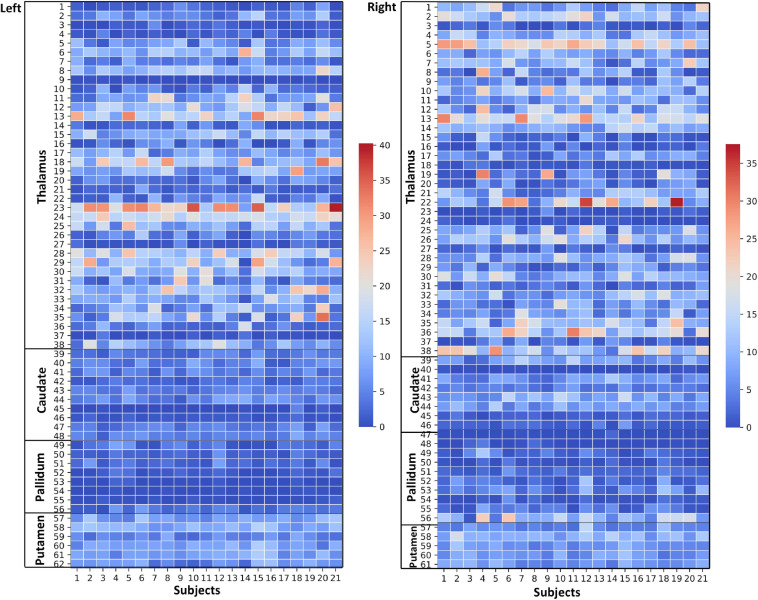
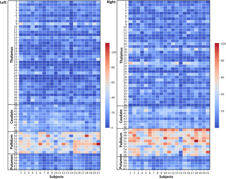
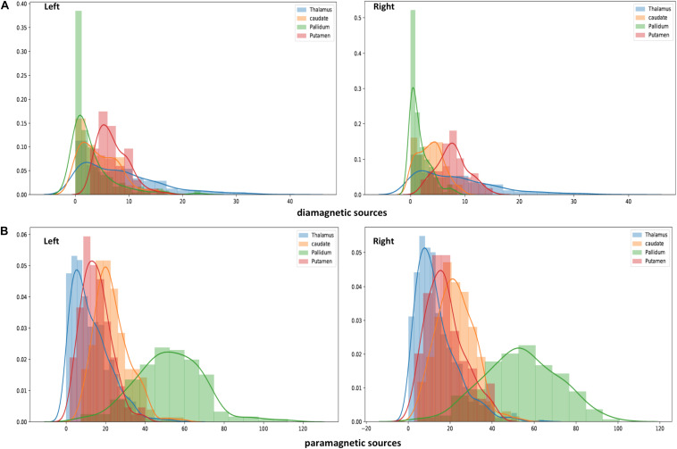
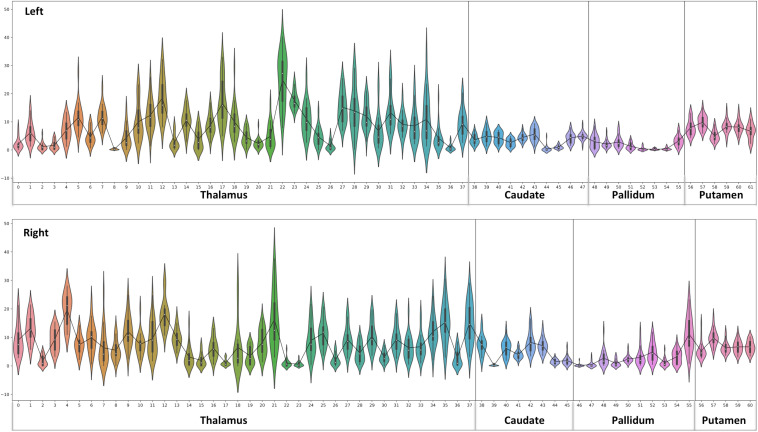
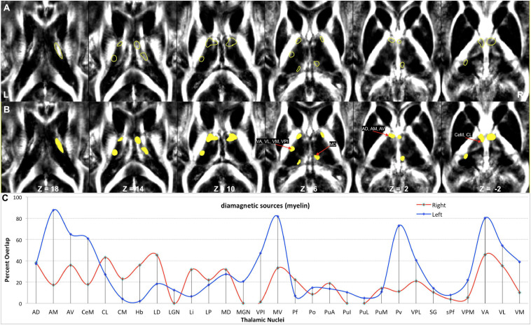
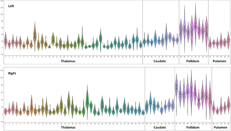
The thalamus (Th) and basal ganglia (BG) are central subcortical connectivity hubs of the human brain, whose functional anatomy is still under intense investigation. Nevertheless, both substructures contain a robust and reproducible functional anatomy. The quantitative susceptibility mapping (QSM) at ultra-high field may facilitate an improved characterization of the underlying functional anatomy . We acquired high-resolution QSM data at 9.4 Tesla in 21 subjects, and analyzed the thalamic and BG by using a prior defined functional parcellation. We found a more substantial contribution of paramagnetic susceptibility sources such as iron in the pallidum in contrast to the caudate, putamen, and Th in descending order. The diamagnetic susceptibility sources such as myelin and calcium revealed significant contributions in the Th parcels compared with the BG. This study presents a detailed nuclei-specific delineation of QSM-provided diamagnetic and paramagnetic susceptibility sources pronounced in the BG and the Th. We also found a reasonable interindividual variability as well as slight hemispheric differences. The results presented here contribute to the microstructural knowledge of the Th and the BG. In specific, the study illustrates QSM values (myelin, calcium, and iron) in functionally similar subregions of the Th and the BG.

摘要

丘脑(Th)和基底神经节(BG)是人类大脑皮层下的中枢连接枢纽,其功能解剖结构仍在深入研究中。然而,这两个亚结构都具有强大且可重复的功能解剖结构。超高场强下的定量磁化率成像(QSM)可能有助于更好地描述其潜在的功能解剖结构。我们在9.4特斯拉场强下采集了21名受试者的高分辨率QSM数据,并使用预先定义的功能分区对丘脑和基底神经节进行了分析。我们发现,与尾状核、壳核和丘脑相比,苍白球中铁等顺磁磁化率源的贡献更大,呈递减顺序。与基底神经节相比,髓磷脂和钙等抗磁磁化率源在丘脑分区中显示出显著贡献。本研究详细描述了QSM提供的在基底神经节和丘脑中明显的抗磁和顺磁磁化率源的核特异性特征。我们还发现了合理的个体间变异性以及轻微的半球差异。这里呈现的结果有助于了解丘脑和基底神经节的微观结构知识。具体而言,该研究展示了丘脑和基底神经节功能相似亚区域的QSM值(髓磷脂、钙和铁)。

https://cdn.ncbi.nlm.nih.gov/pmc/blobs/e156/8483181/bf04cbba250a/fnana-15-725731-g011.jpg
https://cdn.ncbi.nlm.nih.gov/pmc/blobs/e156/8483181/331b6b0d7672/fnana-15-725731-g001.jpg
https://cdn.ncbi.nlm.nih.gov/pmc/blobs/e156/8483181/16d3661abf08/fnana-15-725731-g002.jpg
https://cdn.ncbi.nlm.nih.gov/pmc/blobs/e156/8483181/419beec9e460/fnana-15-725731-g003.jpg
https://cdn.ncbi.nlm.nih.gov/pmc/blobs/e156/8483181/9e5dd9558d14/fnana-15-725731-g004.jpg
https://cdn.ncbi.nlm.nih.gov/pmc/blobs/e156/8483181/f97191894a35/fnana-15-725731-g005.jpg
https://cdn.ncbi.nlm.nih.gov/pmc/blobs/e156/8483181/187ebb13c88f/fnana-15-725731-g006.jpg
https://cdn.ncbi.nlm.nih.gov/pmc/blobs/e156/8483181/ce77f2e15685/fnana-15-725731-g007.jpg
https://cdn.ncbi.nlm.nih.gov/pmc/blobs/e156/8483181/09b6fbfd1cd9/fnana-15-725731-g008.jpg
https://cdn.ncbi.nlm.nih.gov/pmc/blobs/e156/8483181/ad17beff75dc/fnana-15-725731-g009.jpg
https://cdn.ncbi.nlm.nih.gov/pmc/blobs/e156/8483181/7b18cf72e925/fnana-15-725731-g010.jpg
https://cdn.ncbi.nlm.nih.gov/pmc/blobs/e156/8483181/bf04cbba250a/fnana-15-725731-g011.jpg

相似文献

1
Quantitative Susceptibility Mapping of the Basal Ganglia and Thalamus at 9.4 Tesla.9.4特斯拉下基底神经节和丘脑的定量易感性图谱
Front Neuroanat. 2021 Sep 16;15:725731. doi: 10.3389/fnana.2021.725731. eCollection 2021.
2
APART-QSM: An improved sub-voxel quantitative susceptibility mapping for susceptibility source separation using an iterative data fitting method.APART-QSM:一种使用迭代数据拟合方法进行磁化率源分离的改进型亚体素定量磁化率成像。
Neuroimage. 2023 Jul 1;274:120148. doi: 10.1016/j.neuroimage.2023.120148. Epub 2023 Apr 29.
3
Nuclei-specific deposits of iron and calcium in the rat thalamus after status epilepticus revealed with quantitative susceptibility mapping (QSM).癫痫持续状态后大鼠丘脑内铁和钙的核特异性沉积的定量磁化率映射(QSM)显示。
J Magn Reson Imaging. 2018 Feb;47(2):554-564. doi: 10.1002/jmri.25777. Epub 2017 Jun 5.
4
The influence of brain iron and myelin on magnetic susceptibility and effective transverse relaxation - A biochemical and histological validation study.脑铁和髓鞘对磁化率和有效横向弛豫的影响 - 一项生化和组织学验证研究。
Neuroimage. 2018 Oct 1;179:117-133. doi: 10.1016/j.neuroimage.2018.06.007. Epub 2018 Jun 15.
5
Iron quantification in basal ganglia using quantitative susceptibility mapping in a patient with ALS: a case report and literature review.肌萎缩侧索硬化症患者基底节铁定量的定量磁化率成像:病例报告及文献综述
Front Neurosci. 2023 Sep 27;17:1229082. doi: 10.3389/fnins.2023.1229082. eCollection 2023.
6
Toward in vivo histology: a comparison of quantitative susceptibility mapping (QSM) with magnitude-, phase-, and R2*-imaging at ultra-high magnetic field strength.朝着活体组织学迈进:超高场强下定量磁化率映射(QSM)与幅度成像、相位成像和 R2* 成像的比较。
Neuroimage. 2013 Jan 15;65:299-314. doi: 10.1016/j.neuroimage.2012.09.055. Epub 2012 Oct 2.
7
High-resolution characterisation of the aging brain using simultaneous quantitative susceptibility mapping (QSM) and R2* measurements at 7T.在7T磁场下使用同步定量磁化率成像(QSM)和R2*测量对衰老大脑进行高分辨率表征。
Neuroimage. 2016 Sep;138:43-63. doi: 10.1016/j.neuroimage.2016.05.024. Epub 2016 May 13.
8
The association of myelination in the internal capsule with iron deposition in the basal ganglia in macaques: a magnetic resonance imaging study.猕猴内囊髓鞘形成与基底神经节铁沉积的关联:一项磁共振成像研究。
Quant Imaging Med Surg. 2020 Jul;10(7):1526-1539. doi: 10.21037/qims-19-1014.
9
The functional connectivity of intralaminar thalamic nuclei in the human basal ganglia.人类基底神经节中丘脑板内核的功能连接性。
Hum Brain Mapp. 2015 Apr;36(4):1335-47. doi: 10.1002/hbm.22705. Epub 2014 Nov 27.
10
Reproducibility of R * and quantitative susceptibility mapping (QSM) reconstruction methods in the basal ganglia of healthy subjects.健康受试者基底节区R*和定量磁化率成像(QSM)重建方法的可重复性
NMR Biomed. 2017 Apr;30(4). doi: 10.1002/nbm.3491. Epub 2016 Feb 23.

引用本文的文献

1
A roadmap towards standardized neuroimaging approaches for human thalamic nuclei.通向人类丘脑核标准化神经影像学方法的路线图。
Nat Rev Neurosci. 2024 Dec;25(12):792-808. doi: 10.1038/s41583-024-00867-1. Epub 2024 Oct 17.
2
Direct comparison between F-Flortaucipir tau PET and quantitative susceptibility mapping in progressive supranuclear palsy.F-Flortaucipir tau PET 与定量磁化率映射在进行性核上性麻痹中的直接比较。
Neuroimage. 2024 Feb 1;286:120509. doi: 10.1016/j.neuroimage.2024.120509. Epub 2024 Jan 4.

本文引用的文献

1
Alterations and Associations Between Magnetic Susceptibility of the Basal Ganglia and Diffusion Properties in Alzheimer's Disease.阿尔茨海默病中基底节区磁化率与扩散特性之间的改变及关联
Front Neurosci. 2021 Feb 16;15:616163. doi: 10.3389/fnins.2021.616163. eCollection 2021.
2
Iron Deficiency and Iron Excess Differently Affect Dendritic Architecture of Pyramidal Neurons in the Hippocampus of Piglets.缺铁和铁过载对仔猪海马锥体神经元树突形态结构的影响不同。
J Nutr. 2021 Jan 4;151(1):235-244. doi: 10.1093/jn/nxaa326.
3
Neuroimaging of Basal Ganglia in Neurometabolic Diseases in Children.
儿童神经代谢疾病中基底神经节的神经影像学
Brain Sci. 2020 Nov 12;10(11):849. doi: 10.3390/brainsci10110849.
4
The Amsterdam Ultra-high field adult lifespan database (AHEAD): A freely available multimodal 7 Tesla submillimeter magnetic resonance imaging database.阿姆斯特丹超高场成人寿命数据库(AHEAD):一个免费提供的多模态 7 特斯拉亚毫米磁共振成像数据库。
Neuroimage. 2020 Nov 1;221:117200. doi: 10.1016/j.neuroimage.2020.117200. Epub 2020 Aug 1.
5
Hyperactivity of Basal Ganglia in Patients With Parkinson's Disease During Internally Guided Voluntary Movements.帕金森病患者在内部引导的自主运动过程中基底神经节的活动亢进
Front Neurol. 2019 Aug 7;10:847. doi: 10.3389/fneur.2019.00847. eCollection 2019.
6
Evaluation of iron deposition in brain basal ganglia of patients with Parkinson's disease using quantitative susceptibility mapping.利用定量磁化率成像评估帕金森病患者脑基底节区铁沉积情况。
Eur J Radiol Open. 2019 Apr 29;6:169-174. doi: 10.1016/j.ejro.2019.04.005. eCollection 2019.
7
Iron, Myelin, and the Brain: Neuroimaging Meets Neurobiology.铁、髓鞘和大脑:神经影像学与神经生物学的交汇。
Trends Neurosci. 2019 Jun;42(6):384-401. doi: 10.1016/j.tins.2019.03.009. Epub 2019 Apr 29.
8
MRI Changes in the Thalamus and Basal Ganglia of Full-Term Neonates with Perinatal Asphyxia.围产期窒息足月新生儿丘脑和基底神经节的MRI变化
Neonatology. 2018;114(3):253-260. doi: 10.1159/000489159. Epub 2018 Jun 29.
9
In-vivo quantitative structural imaging of the human midbrain and the superior colliculus at 9.4T.在 9.4T 下对人脑中部和上丘进行活体定量结构成像。
Neuroimage. 2018 Aug 15;177:117-128. doi: 10.1016/j.neuroimage.2018.04.071. Epub 2018 May 2.
10
Functional parcellation using time courses of instantaneous connectivity.基于瞬时连通性时间序列的功能分区
Neuroimage. 2018 Apr 15;170:31-40. doi: 10.1016/j.neuroimage.2017.07.027. Epub 2017 Jul 14.