Department of Psychiatry and Psychotherapy, Jena University Hospital, Philosophenweg 3, 07743, Jena, Germany.
Clinical Affective Neuroimaging Laboratory (CANLAB), Leipziger Str. 44, Building 65, 39120, Magdeburg, Germany.
Transl Psychiatry. 2021 Oct 7;11(1):511. doi: 10.1038/s41398-021-01619-w.
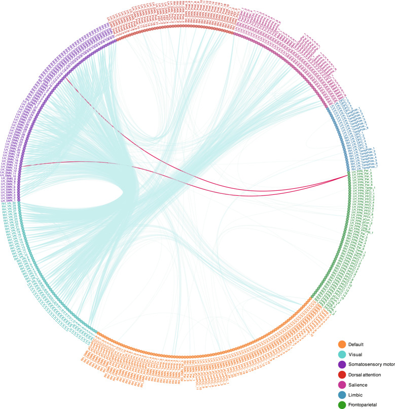
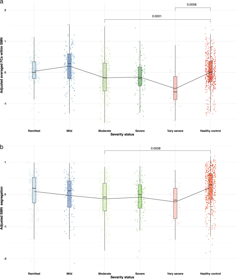
Major depressive disorder (MDD) is associated with abnormal neural circuitry. It can be measured by assessing functional connectivity (FC) at resting-state functional MRI, that may help identifying neural markers of MDD and provide further efficient diagnosis and monitor treatment outcomes. The main aim of the present study is to investigate, in an unbiased way, functional alterations in patients with MDD using a large multi-center dataset from the PsyMRI consortium including 1546 participants from 19 centers ( www.psymri.com ). After applying strict exclusion criteria, the final sample consisted of 606 MDD patients (age: 35.8 ± 11.9 y.o.; females: 60.7%) and 476 healthy participants (age: 33.3 ± 11.0 y.o.; females: 56.7%). We found significant relative hypoconnectivity within somatosensory motor (SMN), salience (SN) networks and between SMN, SN, dorsal attention (DAN), and visual (VN) networks in MDD patients. No significant differences were detected within the default mode (DMN) and frontoparietal networks (FPN). In addition, alterations in network organization were observed in terms of significantly lower network segregation of SMN in MDD patients. Although medicated patients showed significantly lower FC within DMN, FPN, and SN than unmedicated patients, there were no differences between medicated and unmedicated groups in terms of network organization in SMN. We conclude that the network organization of cortical networks, involved in processing of sensory information, might be a more stable neuroimaging marker for MDD than previously assumed alterations in higher-order neural networks like DMN and FPN.
重度抑郁症(MDD)与异常的神经回路有关。可以通过评估静息状态功能磁共振成像的功能连接(FC)来测量,这可能有助于识别 MDD 的神经标志物,并提供进一步有效的诊断和监测治疗效果。本研究的主要目的是使用 PsyMRI 联盟的大型多中心数据集(www.psymri.com),以无偏倚的方式研究 MDD 患者的功能改变,该数据集包括来自 19 个中心的 1546 名参与者。在应用严格的排除标准后,最终样本包括 606 名 MDD 患者(年龄:35.8±11.9 岁;女性:60.7%)和 476 名健康参与者(年龄:33.3±11.0 岁;女性:56.7%)。我们发现 MDD 患者的躯体感觉运动(SMN)、突显(SN)网络内以及 SMN、SN、背侧注意(DAN)和视觉(VN)网络之间的相对连接性降低。在默认模式(DMN)和额顶网络(FPN)内未检测到显著差异。此外,还观察到网络组织的改变,表现为 MDD 患者的 SMN 网络分离程度明显降低。尽管用药患者在 DMN、FPN 和 SN 内的 FC 明显低于未用药患者,但在 SMN 的网络组织方面,用药和未用药组之间没有差异。我们得出结论,参与感觉信息处理的皮质网络的网络组织可能是 MDD 的更稳定的神经影像学标志物,而不是以前认为的 DMN 和 FPN 等高级神经网络的改变。