Department of Oncology, The Second Affiliated Hospital of Nanchang University, Nanchang, Jiangxi 330006, P.R. China.
Department of Oncology, The Second Affiliated Hospital of Nanchang University, Nanchang, Jiangxi 330006, P.R. China.
Mol Med Rep. 2021 Dec;24(6). doi: 10.3892/mmr.2021.12482. Epub 2021 Oct 11.
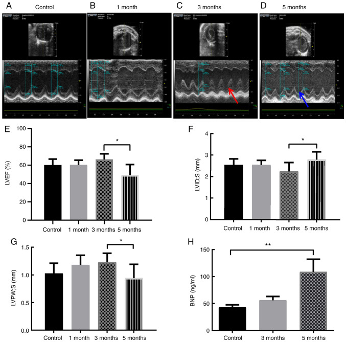
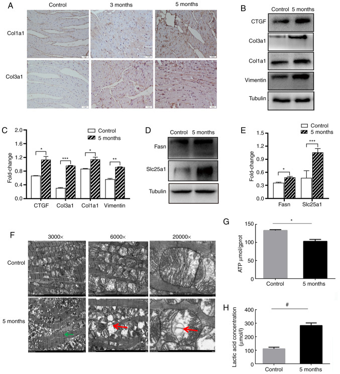
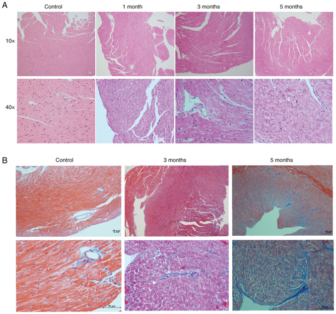
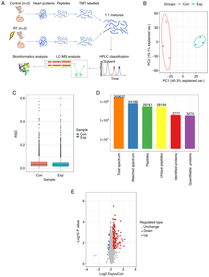
Thoracic radiotherapy increases the risk of radiation‑induced heart damage (RIHD); however, the molecular mechanisms underlying these changes are not fully understood. The aim of the present study was to investigate the effects of radiation on the mouse heart using high‑throughput proteomics. Male C57BL/6J mice were used to establish a model of RIHD by exposing the entire heart to 16 Gy high‑energy X‑rays, and cardiac injuries were verified using a cardiac echocardiogram, as well as by measuring serum brain natriuretic peptide levels and conducting H&E and Masson staining 5 months after irradiation. Proteomics experiments were performed using the heart apex of 5‑month irradiated mice and control mice that underwent sham‑irradiation. The most significantly differentially expressed proteins were enriched in 'cardiac fibrosis' and 'energy metabolism'. Next, the cardiac fibrosis and changes to energy metabolism were confirmed using immunohistochemistry staining and western blotting. Extracellular matrix proteins, such as collagen type 1 α 1 chain, collagen type III α 1 chain, vimentin and CCCTC‑binding factor, along with metabolism‑related proteins, such as fatty acid synthase and solute carrier family 25 member 1, exhibited upregulated expression following exposure to ionizing radiation. Additionally, the myocardial mitochondria inner membranes were injured, along with a decrease in ATP levels and the accumulation of lactic acid in the irradiated heart tissues. These results suggest that the high doses of ionizing radiation used lead to structural remodeling, functional injury and fibrotic alterations in the mouse heart. Radiation‑induced mitochondrial damage and metabolic alterations of the cardiac tissue may thus be a pathogenic mechanism of RIHD.
胸部放射治疗会增加放射性心脏损伤(RIHD)的风险;然而,这些变化的分子机制尚不完全清楚。本研究旨在使用高通量蛋白质组学研究射线对小鼠心脏的影响。雄性 C57BL/6J 小鼠通过暴露整个心脏接受 16 Gy 高能 X 射线来建立 RIHD 模型,并使用心脏超声心动图、测量血清脑钠肽水平以及进行 H&E 和 Masson 染色来验证心脏损伤,照射后 5 个月。使用 5 个月照射的小鼠和接受假照射的对照小鼠的心脏顶端进行蛋白质组学实验。差异最显著的蛋白质在“心脏纤维化”和“能量代谢”中富集。接下来,使用免疫组织化学染色和 Western blot 确认心脏纤维化和能量代谢变化。细胞外基质蛋白,如胶原类型 1α1 链、胶原类型 3α1 链、波形蛋白和 CCCTC 结合因子,以及代谢相关蛋白,如脂肪酸合酶和溶质载体家族 25 成员 1,在暴露于电离辐射后表达上调。此外,心肌线粒体内膜受损,同时照射心脏组织中 ATP 水平降低和乳酸积累。这些结果表明,使用的高剂量电离辐射导致小鼠心脏的结构重塑、功能损伤和纤维化改变。因此,辐射诱导的线粒体损伤和心脏组织的代谢改变可能是 RIHD 的发病机制之一。