Saito Shintaro, Nakajima Kenichi, Edenbrandt Lars, Enqvist Olof, Ulén Johannes, Kinuya Seigo
Department of Nuclear Medicine, Kanazawa University Graduate School of Medicine, 13-1 Takara-machi, Kanazawa, 920-8640, Japan.
Department of Functional Imaging and Artificial Intelligence, Kanazawa University Graduate School of Medicine, 13-1 Takara-machi, Kanazawa, 920-8640, Japan.
EJNMMI Res. 2021 Oct 12;11(1):105. doi: 10.1186/s13550-021-00847-x.
Since three-dimensional segmentation of cardiac region in I-metaiodobenzylguanidine (MIBG) study has not been established, this study aimed to achieve organ segmentation using a convolutional neural network (CNN) with I-MIBG single photon emission computed tomography (SPECT) imaging, to calculate heart counts and washout rates (WR) automatically and to compare with conventional quantitation based on planar imaging.
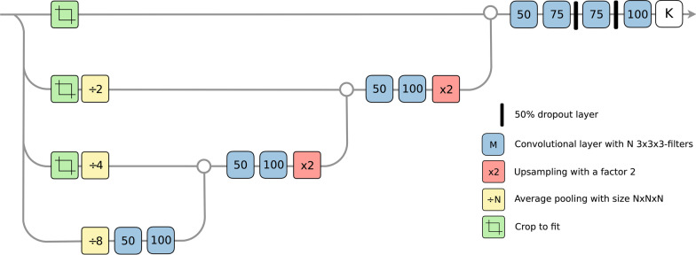
We assessed 48 patients (aged 68.4 ± 11.7 years) with heart and neurological diseases, including chronic heart failure, dementia with Lewy bodies, and Parkinson's disease. All patients were assessed by early and late I-MIBG planar and SPECT imaging. The CNN was initially trained to individually segment the lungs and liver on early and late SPECT images. The segmentation masks were aligned, and then, the CNN was trained to directly segment the heart, and all models were evaluated using fourfold cross-validation. The CNN-based average heart counts and WR were calculated and compared with those determined using planar parameters. The CNN-based SPECT and conventional planar heart counts were corrected by physical time decay, injected dose of I-MIBG, and body weight. We also divided WR into normal and abnormal groups from linear regression lines determined by the relationship between planar WR and CNN-based WR and then analyzed agreement between them.
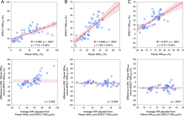
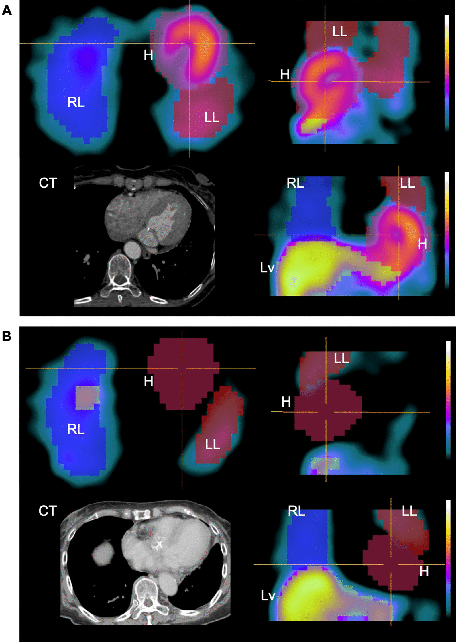
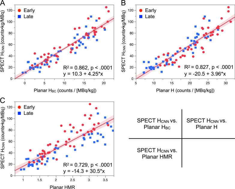
The CNN segmented the cardiac region in patients with normal and reduced uptake. The CNN-based SPECT heart counts significantly correlated with conventional planar heart counts with and without background correction and a planar heart-to-mediastinum ratio (R = 0.862, 0.827, and 0.729, p < 0.0001, respectively). The CNN-based and planar WRs also correlated with and without background correction and WR based on heart-to-mediastinum ratios of R = 0.584, 0.568 and 0.507, respectively (p < 0.0001). Contingency table findings of high and low WR (cutoffs: 34% and 30% for planar and SPECT studies, respectively) showed 87.2% agreement between CNN-based and planar methods.
The CNN could create segmentation from SPECT images, and average heart counts and WR were reliably calculated three-dimensionally, which might be a novel approach to quantifying SPECT images of innervation.
由于123I-间碘苄胍(MIBG)研究中心脏区域的三维分割尚未确立,本研究旨在利用卷积神经网络(CNN)对I-MIBG单光子发射计算机断层扫描(SPECT)成像进行器官分割,自动计算心脏计数和洗脱率(WR),并与基于平面成像的传统定量方法进行比较。
我们评估了48例患有心脏和神经系统疾病的患者(年龄68.4±11.7岁),包括慢性心力衰竭、路易体痴呆和帕金森病。所有患者均接受早期和晚期I-MIBG平面及SPECT成像评估。CNN最初在早期和晚期SPECT图像上分别进行训练,以分割肺和肝脏。将分割掩码对齐后,对CNN进行训练以直接分割心脏,所有模型均采用四折交叉验证进行评估。计算基于CNN的平均心脏计数和WR,并与使用平面参数确定的值进行比较。基于CNN的SPECT和传统平面心脏计数通过物理时间衰减、I-MIBG注射剂量和体重进行校正。我们还根据平面WR与基于CNN的WR之间的关系确定的线性回归线,将WR分为正常组和异常组,然后分析它们之间的一致性。
CNN能够对摄取正常和降低的患者的心脏区域进行分割。基于CNN的SPECT心脏计数与经过和未经过背景校正的传统平面心脏计数以及平面心脏与纵隔比值显著相关(R分别为0.862、0.827和0.729,p均<0.0001)。基于CNN的WR和平面WR在经过和未经过背景校正时也相关,基于心脏与纵隔比值的WR分别为R = 0.584、0.568和0.507(p<0.0001)。高和低WR(截断值:平面研究为34%,SPECT研究为30%)的列联表结果显示,基于CNN的方法与平面方法之间的一致性为87.2%。
CNN能够从SPECT图像创建分割,并可靠地三维计算平均心脏计数和WR,这可能是一种量化神经支配SPECT图像的新方法。