• 文献检索
  • 文档翻译
  • 深度研究
  • 学术资讯
  • Suppr Zotero 插件Zotero 插件
  • 邀请有礼
  • 套餐&价格
  • 历史记录
应用&插件
Suppr Zotero 插件Zotero 插件浏览器插件Mac 客户端Windows 客户端微信小程序
定价
高级版会员购买积分包购买API积分包
服务
文献检索文档翻译深度研究API 文档MCP 服务
关于我们
关于 Suppr公司介绍联系我们用户协议隐私条款
关注我们

Suppr 超能文献

核心技术专利:CN118964589B侵权必究
粤ICP备2023148730 号-1Suppr @ 2026

文献检索

告别复杂PubMed语法,用中文像聊天一样搜索,搜遍4000万医学文献。AI智能推荐,让科研检索更轻松。

立即免费搜索

文件翻译

保留排版,准确专业,支持PDF/Word/PPT等文件格式,支持 12+语言互译。

免费翻译文档

深度研究

AI帮你快速写综述,25分钟生成高质量综述,智能提取关键信息,辅助科研写作。

立即免费体验

7T 与 3T 颅内动脉瘤壁血管壁 MRI 对比评估。

Comparison of 7 T and 3 T vessel wall MRI for the evaluation of intracranial aneurysm wall.

机构信息

Department of Interventional Neuroradiology, Beijing Neurosurgical Institute and Beijing Tiantan Hospital, Capital Medical University, Beijing, China.

State Key Laboratory of Brain and Cognitive Science, Institute of Biophysics, Chinese Academy of Sciences, 15 Datun Road, Beijing, 100101, China.

出版信息

Eur Radiol. 2022 Apr;32(4):2384-2392. doi: 10.1007/s00330-021-08331-9. Epub 2021 Oct 13.

DOI:10.1007/s00330-021-08331-9
PMID:34643780
原文链接:https://pmc.ncbi.nlm.nih.gov/articles/PMC9207191/
Abstract

OBJECTIVES

To compare the visibility of intracranial aneurysm wall and thickness quantification between 7 and 3 T vessel wall imaging and evaluate the association between aneurysm size and wall thickness.

METHODS

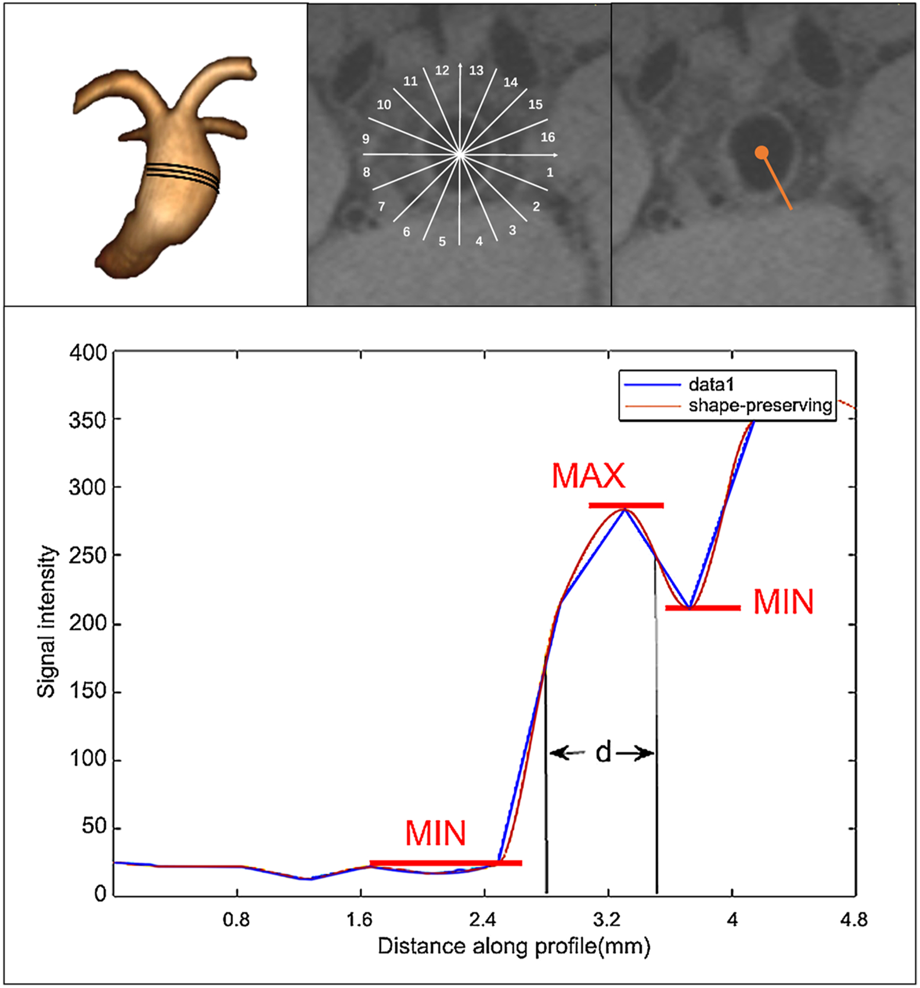
Twenty-nine patients with 29 unruptured intracranial aneurysms were prospectively recruited for 3D T1-weighted vessel wall MRI at both 3 T and 7 T with 0.53 mm (3 T) and 0.4 mm (7 T) isotropic resolution, respectively. Two neuroradiologists independently evaluated wall visibility (0-5 Likert scale), quantified the apparent wall thickness (AWT) using a semi-automated full-width-half-maximum method, calculated wall sharpness, and measured the wall-to-lumen contrast ratio (CR).

RESULTS

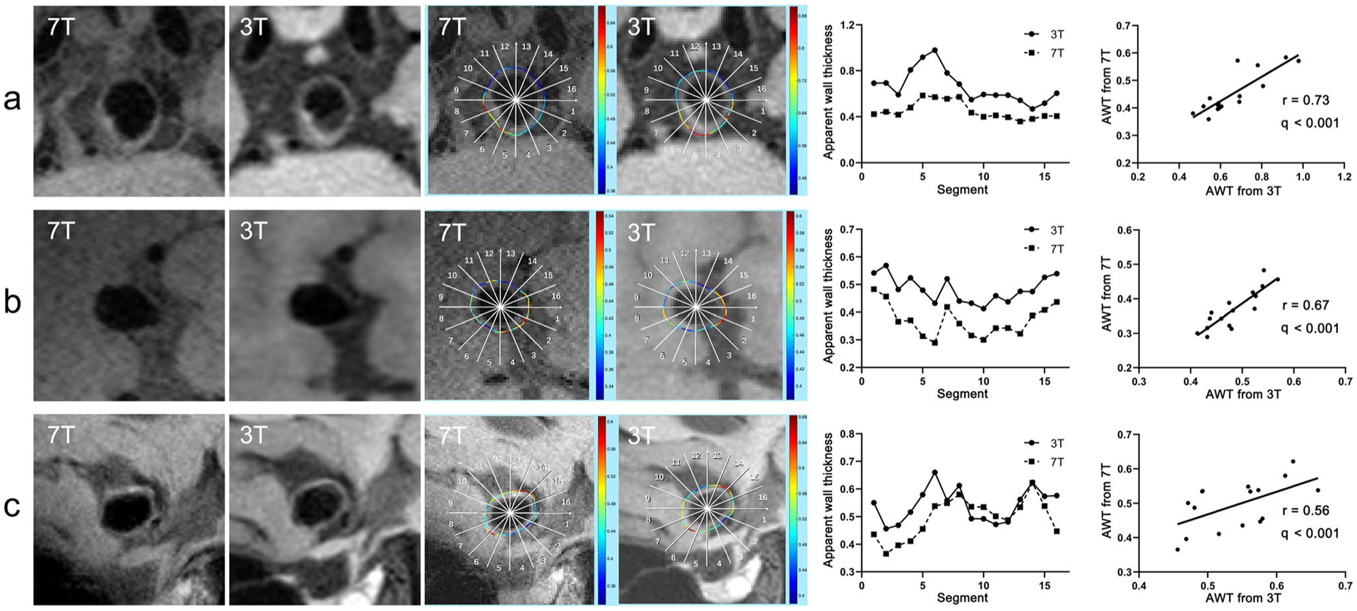
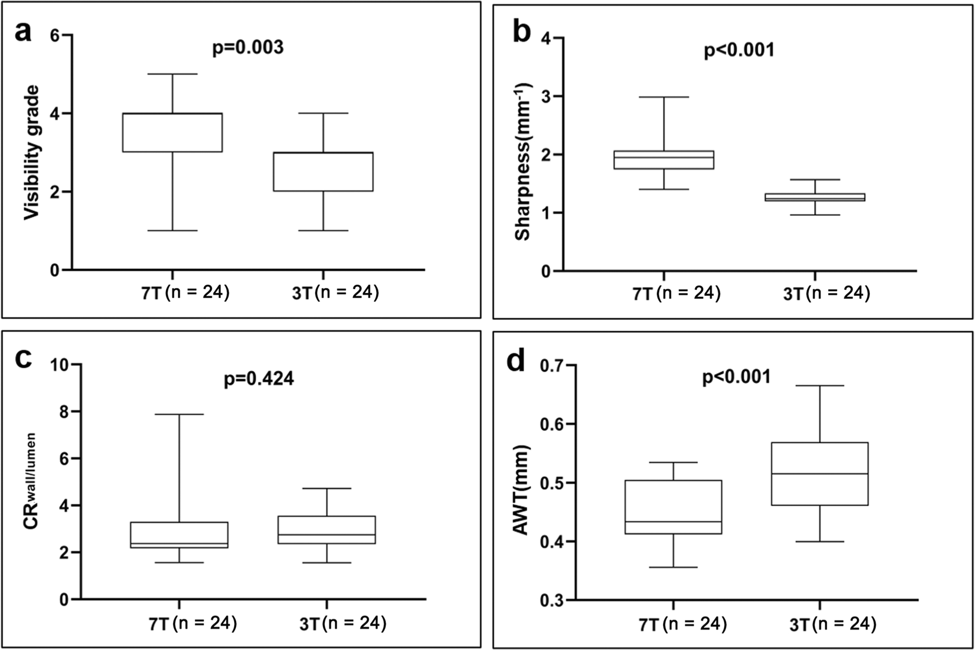
Twenty-four patients with 24 aneurysms were included in this study. 7 T achieved significantly better aneurysm wall visibility than 3 T (3.6 ± 1.1 vs 2.7 ± 0.8, p = 0.003). AWT measured on 3 T and 7 T had a good correlation (averaged r = 0.63 ± 0.19). However, AWT on 3 T was 15% thicker than that on 7 T (0.52 ± 0.07 mm vs 0.45 ± 0.05 mm, p < 0.001). Wall sharpness on 7 T was 57% higher than that on 3 T (1.95 ± 0.32 mm vs 1.24 ± 0.15 mm, p < 0.001). CR on 3 T and 7 T was comparable (p = 0.424). AWT on 7 T was positively correlated with aneurysm size (saccular: r = 0.58, q = 0.046; fusiform: r = 0.67, q = 0.049).

CONCLUSIONS

7 T provides better visualization of intracranial aneurysm wall with higher sharpness than 3 T. 3 T overestimates the wall thickness relative to 7 T. Aneurysm wall thickness is positively correlated with aneurysm size. 7 T MRI is a promising tool to evaluate aneurysm wall in vivo.

KEY POINTS

• 7 T provides better visualization of intracranial aneurysm wall with higher sharpness than 3 T. • 3 T overestimates the wall thickness comparing with 7 T. • Aneurysm wall thickness is positively correlated with aneurysm size.

摘要

目的

比较 7T 和 3T 血管壁成像颅内动脉瘤壁的可视性和厚度定量,并评估动脉瘤大小与壁厚度之间的关系。

方法

前瞻性招募 29 例 29 个未破裂颅内动脉瘤患者,分别在 3T 和 7T 进行 3D T1 加权血管壁 MRI 检查,空间分辨率分别为 0.53mm(3T)和 0.4mm(7T)。两位神经放射科医生独立评估壁的可视性(0-5 级 Likert 量表),使用半自动全宽半高法定量测量表观壁厚度(AWT),计算壁锐度,并测量壁-腔对比度比(CR)。

结果

本研究共纳入 24 例患者的 24 个动脉瘤。7T 比 3T 显著提高了动脉瘤壁的可视性(3.6±1.1 比 2.7±0.8,p=0.003)。3T 和 7T 测量的 AWT 具有良好的相关性(平均 r=0.63±0.19)。然而,3T 上的 AWT 比 7T 厚 15%(0.52±0.07mm 比 0.45±0.05mm,p<0.001)。7T 上的壁锐度比 3T 高 57%(1.95±0.32mm 比 1.24±0.15mm,p<0.001)。3T 和 7T 的 CR 无差异(p=0.424)。7T 上的 AWT 与动脉瘤大小呈正相关(囊状:r=0.58,q=0.046;梭形:r=0.67,q=0.049)。

结论

7T 提供了比 3T 更好的颅内动脉瘤壁可视化效果,壁锐度更高。3T 相对于 7T 高估了壁厚度。动脉瘤壁厚度与动脉瘤大小呈正相关。7T MRI 是一种很有前途的活体评估动脉瘤壁的工具。

https://cdn.ncbi.nlm.nih.gov/pmc/blobs/0777/9207191/9ad13b0851b3/nihms-1816543-f0003.jpg
https://cdn.ncbi.nlm.nih.gov/pmc/blobs/0777/9207191/73a10c37007d/nihms-1816543-f0001.jpg
https://cdn.ncbi.nlm.nih.gov/pmc/blobs/0777/9207191/f6832ce4d1ae/nihms-1816543-f0002.jpg
https://cdn.ncbi.nlm.nih.gov/pmc/blobs/0777/9207191/9ad13b0851b3/nihms-1816543-f0003.jpg
https://cdn.ncbi.nlm.nih.gov/pmc/blobs/0777/9207191/73a10c37007d/nihms-1816543-f0001.jpg
https://cdn.ncbi.nlm.nih.gov/pmc/blobs/0777/9207191/f6832ce4d1ae/nihms-1816543-f0002.jpg
https://cdn.ncbi.nlm.nih.gov/pmc/blobs/0777/9207191/9ad13b0851b3/nihms-1816543-f0003.jpg

相似文献

1
Comparison of 7 T and 3 T vessel wall MRI for the evaluation of intracranial aneurysm wall.7T 与 3T 颅内动脉瘤壁血管壁 MRI 对比评估。
Eur Radiol. 2022 Apr;32(4):2384-2392. doi: 10.1007/s00330-021-08331-9. Epub 2021 Oct 13.
2
Quantitative analysis of unruptured intracranial aneurysm wall thickness and enhancement using 7T high resolution, black blood magnetic resonance imaging.采用 7T 高分辨率、黑血磁共振成像对未破裂颅内动脉瘤壁厚度和增强进行定量分析。
J Neurointerv Surg. 2022 Jul;14(7):723-728. doi: 10.1136/neurintsurg-2021-017688. Epub 2021 Aug 27.
3
High resolution imaging of the intracranial vessel wall at 3 and 7 T using 3D fast spin echo MRI.使用3D快速自旋回波MRI在3T和7T下对颅内血管壁进行高分辨率成像。
MAGMA. 2016 Jun;29(3):559-70. doi: 10.1007/s10334-016-0531-x. Epub 2016 Mar 5.
4
Wall enhancement of intracranial saccular and fusiform aneurysms may differ in intensity and extension: a pilot study using 7-T high-resolution black-blood MRI.颅内囊状和梭形动脉瘤的壁强化在强度和范围上可能有所不同:一项使用 7T 高分辨率黑血 MRI 的初步研究。
Eur Radiol. 2020 Jan;30(1):301-307. doi: 10.1007/s00330-019-06275-9. Epub 2019 Jun 19.
5
Visualizing wall enhancement over time in unruptured intracranial aneurysms using 3D vessel wall imaging.使用三维血管壁成像技术可视化未破裂颅内动脉瘤的壁强化随时间的变化。
J Magn Reson Imaging. 2019 Jul;50(1):193-200. doi: 10.1002/jmri.26553. Epub 2018 Nov 3.
6
Evaluation of cerebral aneurysm wall thickness in experimental aneurysms: comparison of 3T-MR imaging with direct microscopic measurements.实验性动脉瘤中脑动脉瘤壁厚度的评估:3T磁共振成像与直接显微镜测量的比较
Acta Neurochir (Wien). 2014 Jan;156(1):27-34. doi: 10.1007/s00701-013-1919-2. Epub 2013 Oct 30.
7
Complementary Roles of Dynamic Contrast-Enhanced MR Imaging and Postcontrast Vessel Wall Imaging in Detecting High-Risk Intracranial Aneurysms.动态对比增强磁共振成像与增强后血管壁成像在检测高风险颅内动脉瘤中的互补作用。
AJNR Am J Neuroradiol. 2019 Mar;40(3):490-496. doi: 10.3174/ajnr.A5983. Epub 2019 Feb 21.
8
Thinner Regions of Intracranial Aneurysm Wall Correlate with Regions of Higher Wall Shear Stress: A 7T MRI Study.颅内动脉瘤壁较薄区域与较高壁面切应力区域相关:一项7T磁共振成像研究
AJNR Am J Neuroradiol. 2016 Jul;37(7):1310-7. doi: 10.3174/ajnr.A4734. Epub 2016 Feb 18.
9
Visualization of the aneurysm wall: a 7.0-tesla magnetic resonance imaging study.动脉瘤壁的可视化:一项7.0特斯拉磁共振成像研究。
Neurosurgery. 2014 Dec;75(6):614-22; discussion 622. doi: 10.1227/NEU.0000000000000559.
10
Wall enhancement on black-blood MRI is independently associated with symptomatic status of unruptured intracranial saccular aneurysm.黑血 MRI 上的壁强化与未破裂颅内囊状动脉瘤的症状状态独立相关。
Eur Radiol. 2020 Dec;30(12):6413-6420. doi: 10.1007/s00330-020-07063-6. Epub 2020 Jul 14.

引用本文的文献

1
7T MRI in the evaluation of ischemic stroke: a systematic review.7T磁共振成像在缺血性脑卒中评估中的应用:一项系统综述
Front Neurosci. 2025 Jun 23;19:1539617. doi: 10.3389/fnins.2025.1539617. eCollection 2025.
2
Beyond Size: Advanced MRI Breakthroughs in Predicting Intracranial Aneurysm Rupture Risk.超越大小:预测颅内动脉瘤破裂风险的先进磁共振成像突破
J Clin Med. 2025 May 2;14(9):3158. doi: 10.3390/jcm14093158.
3
Abnormal changes in neuropsychological function, brain structure and cerebral perfusion in patients with unruptured intracranial aneurysms.

本文引用的文献

1
Evaluation of direct-to-angiography suite (DTAS) and conventional clinical pathways in stroke care: a simulation study.直接到导管室(DTAS)和传统临床路径在卒中护理中的评估:一项模拟研究。
J Neurointerv Surg. 2022 Dec;14(12):1189-1194. doi: 10.1136/neurintsurg-2021-018253. Epub 2021 Dec 6.
2
Decreased contrast enhancement on high-resolution vessel wall imaging of unruptured intracranial aneurysms in patients taking aspirin.服用阿司匹林的未破裂颅内动脉瘤患者的高分辨率血管壁成像的对比增强减弱。
J Neurosurg. 2020 Mar 6;134(3):902-908. doi: 10.3171/2019.12.JNS193023. Print 2021 Mar 1.
3
Test-retest multisite reproducibility of neurovascular 4D flow MRI.
未破裂颅内动脉瘤患者神经心理功能、脑结构和脑灌注的异常变化。
Front Neurol. 2024 Oct 8;15:1463156. doi: 10.3389/fneur.2024.1463156. eCollection 2024.
4
Occult Amyloid-β-Related Angiitis: Neuroimaging Findings at 1.5T, 3T, and 7T MRI.隐匿性β淀粉样蛋白相关血管炎:1.5T、3T 和 7T MRI 的神经影像学表现。
AJNR Am J Neuroradiol. 2024 Aug 9;45(8):1013-1018. doi: 10.3174/ajnr.A8264.
5
Diagnosis of Unruptured Intracranial Aneurysms Using Proton-Density Magnetic Resonance Angiography: A Comparison With High-Resolution Time-of-Flight Magnetic Resonance Angiography.质子密度磁共振血管造影诊断未破裂颅内动脉瘤:与高分辨率时间飞跃磁共振血管造影的比较。
Korean J Radiol. 2024 Jun;25(6):575-588. doi: 10.3348/kjr.2023.1241.
6
Use of a Commercial 7-T MRI Scanner for Clinical Brain Imaging: Indications, Protocols, Challenges, and Solutions-A Single-Center Experience.商用 7T MRI 扫描仪在临床脑部成像中的应用:单中心经验分享——适应证、方案、挑战及解决方案。
AJR Am J Roentgenol. 2023 Dec;221(6):788-804. doi: 10.2214/AJR.23.29342. Epub 2023 Jun 28.
7
Advanced cross-sectional imaging of cerebral aneurysms.脑动脉瘤的高级影像学检查
Br J Radiol. 2023 Jan 1;96(1141):20220686. doi: 10.1259/bjr.20220686. Epub 2022 Dec 9.
8
Correlation Between Thrombus Signal Intensity and Aneurysm Wall Thickness in Partially Thrombosed Intracranial Aneurysms Using 7T Magnetization-Prepared Rapid Acquisition Gradient Echo Magnetic Resonance Imaging.使用7T磁化准备快速采集梯度回波磁共振成像技术对部分血栓形成的颅内动脉瘤中血栓信号强度与动脉瘤壁厚度之间的相关性研究
Front Neurol. 2022 Feb 18;13:758126. doi: 10.3389/fneur.2022.758126. eCollection 2022.
神经血管 4D 流 MRI 的测试-重测多站点再现性。
J Magn Reson Imaging. 2019 Jun;49(6):1543-1552. doi: 10.1002/jmri.26564. Epub 2018 Nov 15.
4
Growth and rupture of unruptured cerebral aneurysms based on the intraoperative appearance.基于术中表现的未破裂脑动脉瘤的生长与破裂情况
Acta Med Okayama. 1994 Oct;48(5):257-62. doi: 10.18926/AMO/31120.