Department of Cardiology, Henan Provincial People's Hospital, Zhengzhou University People's Hospital, Zhengzhou, Henan, China (mainland).
Department of Radiology, Clinical Medical College and The First Affiliated Hospital of Chengdu Medical College, Chengdu, Sichuan, China (mainland).
Med Sci Monit. 2021 Oct 20;27:e933220. doi: 10.12659/MSM.933220.
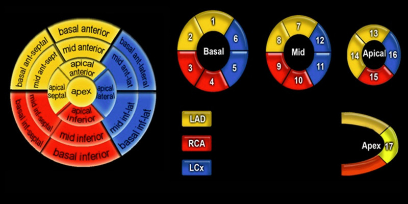
BACKGROUND In this study, cardiac magnetic resonance imaging was used to investigate the characteristics of patients who have total coronary occlusion but manifest with non-ST-segment elevation myocardial infarction (NSTEMI), and we assessed the extent of infarct transmurality and myocardial necrosis size in NSTEMI patients. MATERIAL AND METHODS We enrolled all patients diagnosed at our hospital with subtotal or total occlusion of the culprit artery (TOCA), based on the coronary angiography, who successfully underwent PCI within 12 h of admission, and who had CMR imaging performed within 2 days after the PCI. RESULTS Based on 12-lead ECG findings, 48% of patients were categorized as having STEMI and 52% as having NSTEMI. TOCA was detected by coronary angiography in 43% of NSTEMI patients, and in 60% and 33% of normal ST segment and ST-segment depression MI patients, respectively. The transmural segments were found in 78% of STEMI patients and 31% of NSTEMI patients (P<0.05). Transmural infarction segments were found in 64% of NSTEMI patients with TOCA and in 8% of NTOCA patients (P<0.05). Moreover, the number of transmural segments in ST-segment depression MI patients was the lowest (P<0.05). Infarct size in STEMI patients was significantly larger than in patients with NSTEMI (P<0.05), whereas there was no statistically significant difference in patients with normal ST segment and ST-segment depression MI patients (P>0.05). CONCLUSIONS Identification TOCA by coronary angiography and transmural infarction by DE-MRI can be challenging in AMI patients with non-ST-segment elevation. In approximately 30% of non-ST-segment elevation MI patients, transmural infarction was detected by DE-MRI. Therefore, TOCA accompanied by transmural infarction in non-ST-segment-elevation MI patients is not uncommon.
在这项研究中,我们使用心脏磁共振成像来研究完全性冠状动脉闭塞但表现为非 ST 段抬高型心肌梗死(NSTEMI)的患者的特征,并评估 NSTEMI 患者的梗死透壁程度和心肌坏死面积。
我们纳入了所有因罪犯动脉次全或完全闭塞(TOCA)在我院接受诊断、在入院后 12 小时内成功接受 PCI 且在 PCI 后 2 天内行 CMR 成像的患者。
根据 12 导联心电图结果,48%的患者被归类为 ST 段抬高型心肌梗死,52%的患者被归类为 NSTEMI。TOCA 在 43%的 NSTEMI 患者中通过冠状动脉造影检测到,在正常 ST 段和 ST 段压低 MI 患者中分别为 60%和 33%。78%的 ST 段抬高型心肌梗死患者和 31%的 NSTEMI 患者存在透壁节段(P<0.05)。TOCA 组的 NSTEMI 患者中有 64%存在透壁梗死节段,而无 TOCA 的 NSTEMI 患者中仅有 8%存在透壁梗死节段(P<0.05)。此外,ST 段压低 MI 患者的透壁节段数量最少(P<0.05)。ST 段抬高型心肌梗死患者的梗死面积明显大于 NSTEMI 患者(P<0.05),而在正常 ST 段和 ST 段压低 MI 患者中则无统计学差异(P>0.05)。
在非 ST 段抬高型心肌梗死患者中,通过冠状动脉造影识别 TOCA 和通过 DE-MRI 识别透壁梗死具有挑战性。在大约 30%的非 ST 段抬高型心肌梗死患者中,DE-MRI 检测到透壁梗死。因此,非 ST 段抬高型心肌梗死患者中伴有透壁梗死的 TOCA 并不少见。