Department of Synaptic Plasticity, Max Planck Institut fur Hirnforschung, Frankfurt am Main, Hessen 60438, Germany.
Department of Synaptic Plasticity, Max Planck Institut fur Hirnforschung, Frankfurt am Main, Hessen 60438, Germany
Proc Natl Acad Sci U S A. 2021 Oct 26;118(43). doi: 10.1073/pnas.2113929118.
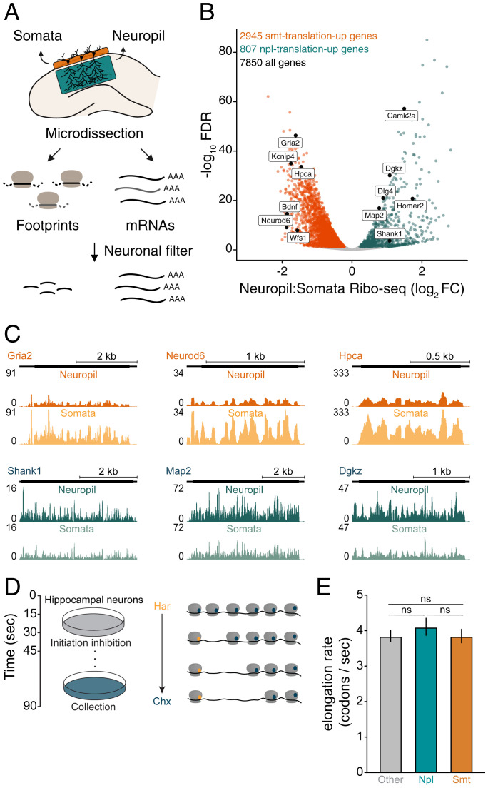
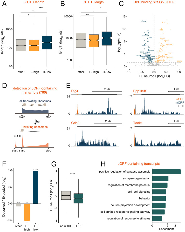
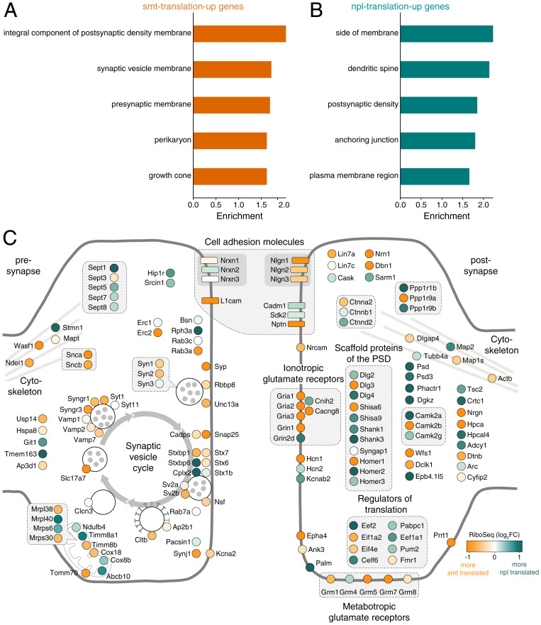
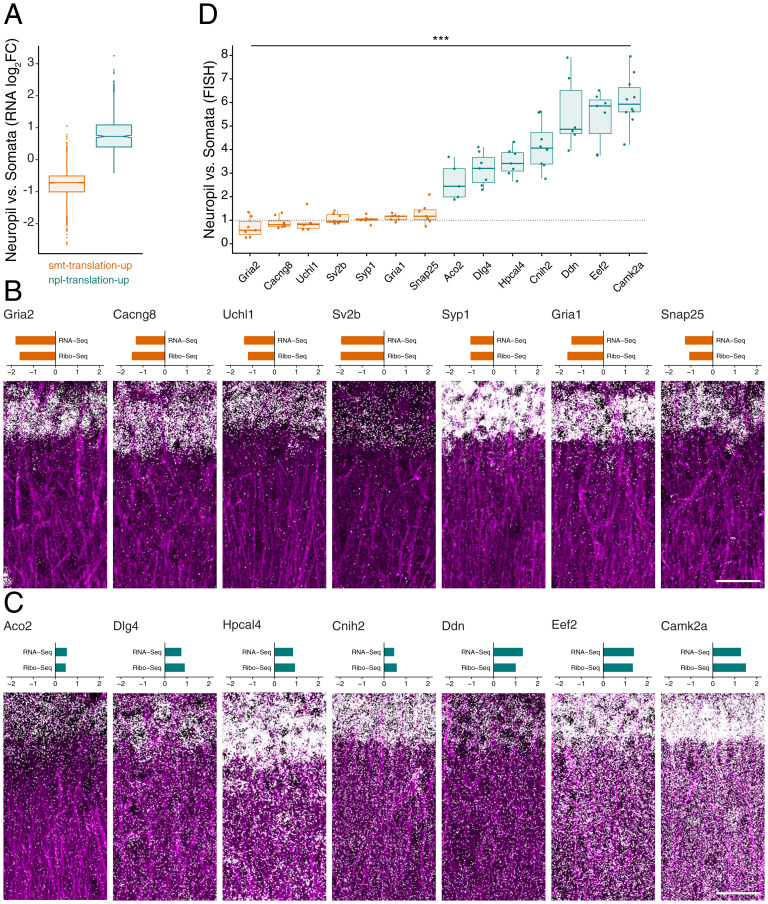
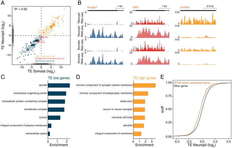
To form synaptic connections and store information, neurons continuously remodel their proteomes. The impressive length of dendrites and axons imposes logistical challenges to maintain synaptic proteins at locations remote from the transcription source (the nucleus). The discovery of thousands of messenger RNAs (mRNAs) near synapses suggested that neurons overcome distance and gain autonomy by producing proteins locally. It is not generally known, however, if, how, and when localized mRNAs are translated into protein. To investigate the translational landscape in neuronal subregions, we performed simultaneous RNA sequencing (RNA-seq) and ribosome sequencing (Ribo-seq) from microdissected rodent brain slices to identify and quantify the transcriptome and translatome in cell bodies (somata) as well as dendrites and axons (neuropil). Thousands of transcripts were differentially translated between somatic and synaptic regions, with many scaffold and signaling molecules displaying increased translation levels in the neuropil. Most translational changes between compartments could be accounted for by differences in RNA abundance. Pervasive translational regulation was observed in both somata and neuropil influenced by specific mRNA features (e.g., untranslated region [UTR] length, RNA-binding protein [RBP] motifs, and upstream open reading frames [uORFs]). For over 800 mRNAs, the dominant source of translation was the neuropil. We constructed a searchable and interactive database for exploring mRNA transcripts and their translation levels in the somata and neuropil [MPI Brain Research, The mRNA translation landscape in the synaptic neuropil. https://public.brain.mpg.de/dashapps/localseq/ Accessed 5 October 2021]. Overall, our findings emphasize the substantial contribution of local translation to maintaining synaptic protein levels and indicate that on-site translational control is an important mechanism to control synaptic strength.
为了形成突触连接和储存信息,神经元不断重塑其蛋白质组。树突和轴突的惊人长度给维持远离转录源(细胞核)的突触蛋白带来了后勤挑战。在突触附近发现数千种信使 RNA(mRNA)表明,神经元通过局部产生蛋白质来克服距离并获得自主性。然而,目前尚不清楚局部 mRNA 是否以及何时被翻译成蛋白质,以及如何翻译。为了研究神经元亚区的翻译景观,我们从微切割的啮齿动物脑片中同时进行 RNA 测序(RNA-seq)和核糖体测序(Ribo-seq),以鉴定和定量细胞体(体)以及树突和轴突(神经胶质)中的转录组和翻译组。数千个转录本在体细胞和突触区之间差异翻译,许多支架和信号分子在神经胶质中的翻译水平增加。大多数隔室之间的翻译变化可以用 RNA 丰度的差异来解释。在躯体和神经胶质中都观察到普遍的翻译调节,受特定 mRNA 特征(例如,非翻译区 [UTR] 长度、RNA 结合蛋白 [RBP] 基序和上游开放阅读框 [uORF])的影响。对于超过 800 个 mRNA,翻译的主要来源是神经胶质。我们构建了一个可搜索和交互式数据库,用于探索体细胞和神经胶质中的 mRNA 转录本及其翻译水平 [MPI 大脑研究,突触神经胶质中的 mRNA 翻译景观。https://public.brain.mpg.de/dashapps/localseq/ 访问日期:2021 年 10 月 5 日]。总体而言,我们的研究结果强调了局部翻译对维持突触蛋白水平的重要贡献,并表明原位翻译控制是控制突触强度的重要机制。