Department of Ophthalmology, Harvard Medical School, Boston Children's Hospital, Boston, MA, USA.
Department of Cardiology, Harvard Medical School, Boston Children's Hospital, Boston, MA, USA.
EBioMedicine. 2021 Nov;73:103632. doi: 10.1016/j.ebiom.2021.103632. Epub 2021 Oct 21.
Pathological neovascularization in neovascular age-related macular degeneration (nAMD) is the leading cause of vision loss in the elderly. Increasing evidence shows that cells of myeloid lineage play important roles in controlling pathological endothelium formation. Suppressor of cytokine signaling 3 (SOCS3) pathway has been linked to neovascularization.
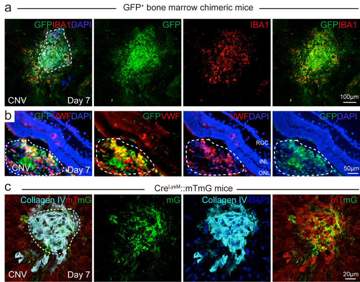
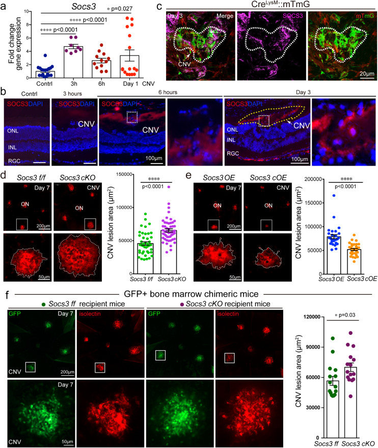
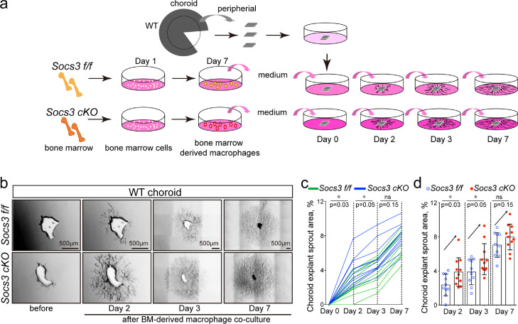
We utilised a laser-induced choroidal neovascularization (CNV) mouse model to investigate the neovascular aspect of human AMD. In several cell lineage reporter mice, bone marrow chimeric mice and Socs3 loss-of-function (knockout) and gain-of-function (overexpression) mice, immunohistochemistry, confocal, and choroidal explant co-culture with bone marrow-derived macrophage medium were used to study the mechanisms underlying pathological CNV formation via myeloid SOCS3.
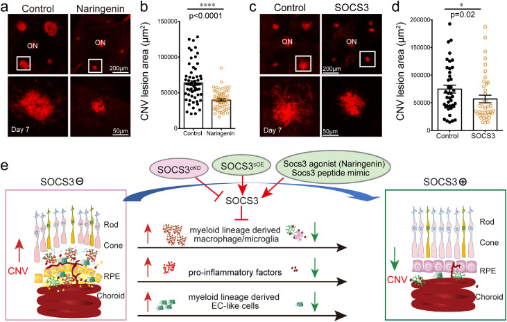
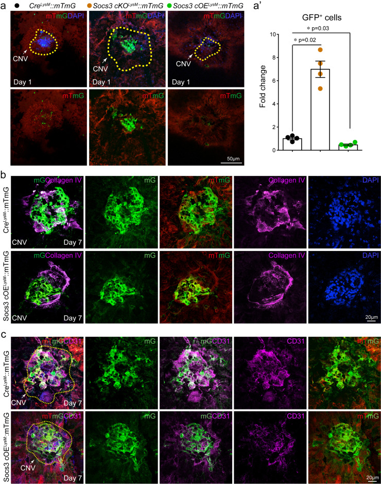
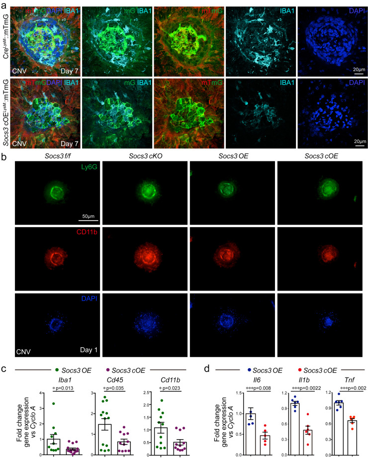
SOCS3 was significantly induced in myeloid lineage cells, which were recruited into the CNV lesion area. Myeloid Socs3 overexpression inhibited laser-induced CNV, reduced myeloid lineage-derived macrophage/microglia recruitment onsite, and attenuated pro-inflammatory factor expression. Moreover, SOCS3 in myeloid regulated vascular sprouting ex vivo in choroid explants and SOCS3 agonist reduced in vivo CNV.
These findings suggest that myeloid lineage cells contributed to pathological CNV formation regulated by SOCS3.
This project was funded by NIH/NEI (R01EY030140, R01EY029238), BrightFocus Foundation, American Health Assistance Foundation (AHAF), and Boston Children's Hospital Ophthalmology Foundation for YS and the National Institutes of Health/National Heart, Lung and Blood Institute (U01HL098166) for PZ.
新生血管性年龄相关性黄斑变性(nAMD)中的病理性血管新生是老年人视力丧失的主要原因。越来越多的证据表明,髓系细胞在控制病理性血管内皮形成中发挥重要作用。细胞因子信号转导抑制因子 3(SOCS3)途径与血管新生有关。
我们利用激光诱导脉络膜新生血管(CNV)小鼠模型来研究人类 AMD 的新生血管方面。在几种细胞谱系报告小鼠、骨髓嵌合小鼠以及 Socs3 功能丧失(敲除)和功能获得(过表达)小鼠中,我们通过免疫组织化学、共聚焦显微镜和脉络膜外植体与骨髓来源的巨噬细胞培养基的共培养,研究了通过髓系 SOCS3 导致病理性 CNV 形成的机制。
SOCS3 在髓系细胞中被显著诱导,这些细胞被募集到 CNV 病变区域。髓系 Socs3 过表达抑制激光诱导的 CNV,减少了骨髓源性巨噬细胞/小胶质细胞在原位的募集,并减弱了促炎因子的表达。此外,髓系中的 SOCS3 调节脉络膜外植体中的血管发芽,SOCS3 激动剂减少体内 CNV。
这些发现表明,髓系细胞有助于 SOCS3 调节的病理性 CNV 形成。
这项研究由美国国立卫生研究院/国家眼科研究所(R01EY030140、R01EY029238)、美国国家眼科研究所、美国心脏协会基金会(AHAF)和波士顿儿童医院眼科基金会资助(AHAF)和波士顿儿童医院眼科基金会为 YS 提供资金,美国国立卫生研究院/美国国立心肺血液研究所(U01HL098166)为 PZ 提供资金。