Department of Diagnostic and Interventional Radiology, Osaka International Cancer Institute, 3-1-69 Otemae, Chuo-ku, Osaka, 541-8567, Japan.
Department of Radiation Oncology, Osaka International Cancer Institute, 3-1-69, Otemae, Chuo-ku, Osaka, 541-8567, Japan.
Jpn J Radiol. 2022 Mar;40(3):229-244. doi: 10.1007/s11604-021-01205-6. Epub 2021 Oct 25.
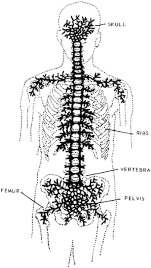
Whole-body magnetic resonance imaging (WB-MRI) is currently used worldwide for detecting bone metastases from prostate cancer. The 5-year survival rate for prostate cancer is > 95%. However, an increase in survival time may increase the incidence of bone metastasis. Therefore, detecting bone metastases is of great clinical interest. Bone metastases are commonly located in the spine, pelvis, shoulder, and distal femur. Bone metastases from prostate cancer are well-known representatives of osteoblastic metastases. However, other types of bone metastases, such as mixed or inter-trabecular type, have also been detected using MRI. MRI does not involve radiation exposure and has good sensitivity and specificity for detecting bone metastases. WB-MRI has undergone gradual developments since the last century, and in 2004, Takahara et al., developed diffusion-weighted Imaging (DWI) with background body signal suppression (DWIBS). Since then, WB-MRI, including DWI, has continued to play an important role in detecting bone metastases and monitoring therapeutic effects. An imaging protocol that allows complete examination within approximately 30 min has been established. This review focuses on WB-MRI standardization and the automatic calculation of tumor total diffusion volume (tDV) and mean apparent diffusion coefficient (ADC) value. In the future, artificial intelligence (AI) will enable shorter imaging times and easier automatic segmentation.
全身磁共振成像(WB-MRI)目前在全球范围内用于检测前列腺癌的骨转移。前列腺癌的 5 年生存率超过 95%。然而,生存时间的延长可能会增加骨转移的发生率。因此,检测骨转移具有重要的临床意义。骨转移通常位于脊柱、骨盆、肩部和股骨远端。前列腺癌的骨转移是成骨性转移的典型代表。然而,其他类型的骨转移,如混合性或小梁内型,也已通过 MRI 检测到。MRI 不涉及辐射,对检测骨转移具有良好的敏感性和特异性。自上个世纪以来,WB-MRI 经历了逐步发展,2004 年,Takahara 等人开发了背景抑制的弥散加权成像(DWIBS)。此后,包括 DWI 在内的 WB-MRI 继续在检测骨转移和监测治疗效果方面发挥重要作用。已经建立了一种允许在大约 30 分钟内完成全面检查的成像方案。本综述重点介绍 WB-MRI 的标准化和肿瘤总弥散体积(tDV)和平均表观弥散系数(ADC)值的自动计算。未来,人工智能(AI)将实现更短的成像时间和更简单的自动分割。