Vascular Cognitive Impairment and Neurodegeneration Program, Oklahoma Center for Geroscience and Healthy Brain Aging, Department of Biochemistry and Molecular Biology, University of Oklahoma Health Sciences Center, 975 NE 10th Street, BRC 1301, Oklahoma City, OK, 73104, USA.
Division of Clinical Physiology, Department of Cardiology, Faculty of Medicine, University of Debrecen, Debrecen, Hungary.
Sci Rep. 2021 Oct 25;11(1):20994. doi: 10.1038/s41598-021-00188-8.
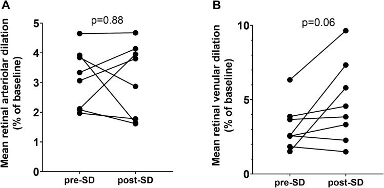
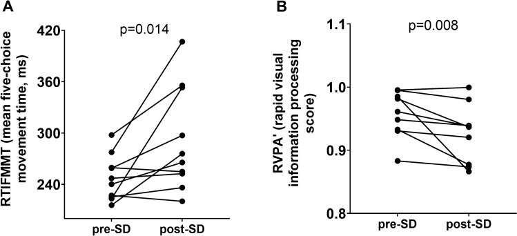
Sleep deprivation (SD) is a common condition and an important health concern. In addition to metabolic and cardiovascular risks, SD associates with decreases in cognitive performance. Neurovascular coupling (NVC, "functional hyperemia") is a critical homeostatic mechanism, which maintains adequate blood supply to the brain during periods of intensive neuronal activity. To determine whether SD alters NVC responses and cognitive performance, cognitive and hemodynamic NVC assessments were conducted prior to and 24 h post-SD in healthy young male individuals (n = 10, 27 ± 3 years old). Cognition was evaluated with a battery of tests from the Cambridge Neuropsychological Test Automated Battery (CANTAB). Hemodynamic components of NVC were measured by transcranial Doppler sonography (TCD) during cognitive stimulation, dynamic retinal vessel analysis (DVA) during flicker light stimulation, and functional near infrared spectroscopy (fNIRS) during finger tapping motor task. Cognitive assessments revealed impairments in reaction time and sustained attention after 24 h of SD. Functional NIRS analysis revealed that SD significantly altered hemodynamic responses in the prefrontal cortex and somatosensory cortex during a motor task. NVC-related vascular responses measured by DVA and TCD did not change significantly. Interestingly, TCD detected decreased task-associated cerebral blood flow (CBF) in the right middle cerebral artery in sleep deprived participants. Our results demonstrate that 24 h of SD lead to impairments in cognitive performance together with altered CBF and hemodynamic components of cortical NVC responses.
睡眠剥夺(SD)是一种常见的状况,也是一个重要的健康问题。除了代谢和心血管风险外,SD 还与认知表现下降有关。神经血管耦合(NVC,“功能充血”)是一种关键的体内平衡机制,可在神经元活动密集期间维持大脑的充足血液供应。为了确定 SD 是否改变了 NVC 反应和认知表现,在健康年轻男性个体(n=10,27±3 岁)进行 SD 前和 24 小时后进行了认知和血液动力学 NVC 评估。认知评估使用剑桥神经心理学测试自动化电池(CANTAB)中的一系列测试进行。在认知刺激期间通过经颅多普勒超声(TCD)测量 NVC 的血液动力学成分,在闪烁光刺激期间通过动态视网膜血管分析(DVA)测量,在手指敲击运动任务期间通过功能近红外光谱(fNIRS)测量。认知评估显示,24 小时 SD 后反应时间和持续注意力受损。功能 fNIRS 分析显示,SD 显著改变了运动任务期间前额叶皮层和体感皮层的血液动力学反应。通过 DVA 和 TCD 测量的与 NVC 相关的血管反应没有明显变化。有趣的是,TCD 检测到睡眠剥夺参与者右侧大脑中动脉的任务相关脑血流(CBF)减少。我们的结果表明,24 小时 SD 导致认知表现受损,同时还改变了 CBF 和皮质 NVC 反应的血液动力学成分。