The Roskamp Institute, 2040 Whitfield Avenue, Sarasota, FL, 34243, USA.
The Open University, Milton Keynes, UK.
Fluids Barriers CNS. 2021 Oct 26;18(1):48. doi: 10.1186/s12987-021-00283-y.
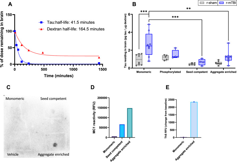
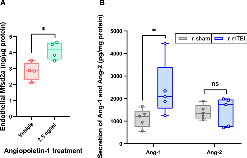
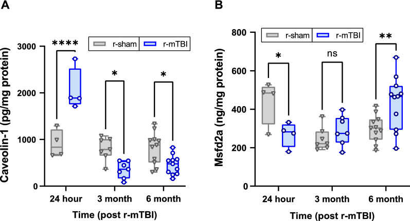
Repetitive head trauma has been associated with the accumulation of tau species in the brain. Our prior work showed brain vascular mural cells contribute to tau processing in the brain, and that these cells progressively degenerate following repetitive mild traumatic brain injury (r-mTBI). The current studies investigated the role of the cerebrovasculature in the elimination of extracellular tau from the brain, and the influence of r-mTBI on these processes. Following intracranial injection of biotin-labeled tau, the levels of exogenous labeled tau residing in the brain were elevated in a mouse model of r-mTBI at 12 months post-injury compared to r-sham mice, indicating reduced tau elimination from the brain following head trauma. This may be the result of decreased caveolin-1 mediated tau efflux at the blood-brain barrier (BBB), as the caveolin inhibitor, methyl-β-cyclodextrin, significantly reduced tau uptake in isolated cerebrovessels and significantly decreased the basolateral-to-apical transit of tau across an in vitro model of the BBB. Moreover, we found that the upstream regulator of endothelial caveolin-1, Mfsd2a, was elevated in r-mTBI cerebrovessels compared to r-sham, which coincided with a decreased expression of cerebrovascular caveolin-1 in the chronic phase following r-mTBI (> 3 months post-injury). Lastly, angiopoietin-1, a mural cell-derived protein governing endothelial Mfsd2a expression, was secreted from r-mTBI cerebrovessels to a greater extent than r-sham animals. Altogether, in the chronic phase post-injury, release of angiopoietin-1 from degenerating mural cells downregulates caveolin-1 expression in brain endothelia, resulting in decreased tau elimination across the BBB, which may describe the accumulation of tau species in the brain following head trauma.
反复的头部创伤与脑中 tau 种的积累有关。我们之前的工作表明,脑血管壁细胞有助于脑中 tau 的处理,并且这些细胞在反复轻度创伤性脑损伤(r-mTBI)后逐渐退化。目前的研究调查了脑血管在从大脑中清除细胞外 tau 中的作用,以及 r-mTBI 对这些过程的影响。在颅内注射生物素标记的 tau 后,与 r-sham 小鼠相比,r-mTBI 小鼠在损伤后 12 个月时脑中存在的外源性标记 tau 水平升高,表明头部创伤后大脑中 tau 的消除减少。这可能是由于血脑屏障(BBB)中 caveolin-1 介导的 tau 外排减少所致,因为 caveolin 抑制剂甲基-β-环糊精可显著减少分离的脑血管中 tau 的摄取,并显著降低 tau 在体外 BBB 模型中的基底外侧至顶端转运。此外,我们发现 r-mTBI 脑血管中的内皮细胞 caveolin-1 的上游调节因子 Mfsd2a 与 r-sham 相比升高,这与 r-mTBI 后慢性期(>损伤后 3 个月)脑血管 caveolin-1 的表达降低相吻合。最后,血管生成素-1,一种调节内皮细胞 Mfsd2a 表达的壁细胞衍生蛋白,从 r-mTBI 脑血管中释放的程度大于 r-sham 动物。总之,在损伤后慢性期,退化的壁细胞从 angiopoietin-1 中释放出来,下调脑内皮细胞中 caveolin-1 的表达,导致 BBB 中 tau 的清除减少,这可能描述了头部创伤后脑中 tau 种的积累。