• 文献检索
  • 文档翻译
  • 深度研究
  • 学术资讯
  • Suppr Zotero 插件Zotero 插件
  • 邀请有礼
  • 套餐&价格
  • 历史记录
应用&插件
Suppr Zotero 插件Zotero 插件浏览器插件Mac 客户端Windows 客户端微信小程序
定价
高级版会员购买积分包购买API积分包
服务
文献检索文档翻译深度研究API 文档MCP 服务
关于我们
关于 Suppr公司介绍联系我们用户协议隐私条款
关注我们

Suppr 超能文献

核心技术专利:CN118964589B侵权必究
粤ICP备2023148730 号-1Suppr @ 2026

文献检索

告别复杂PubMed语法,用中文像聊天一样搜索,搜遍4000万医学文献。AI智能推荐,让科研检索更轻松。

立即免费搜索

文件翻译

保留排版,准确专业,支持PDF/Word/PPT等文件格式,支持 12+语言互译。

免费翻译文档

深度研究

AI帮你快速写综述,25分钟生成高质量综述,智能提取关键信息,辅助科研写作。

立即免费体验

一例伪装成冠状动脉瘤的左心耳病例。

A Case of a Left Atrial Appendage Disguised as a Coronary Artery Aneurysm.

作者信息

Das Nikkan, Tadros Sameh S, DeBrunner Mark

机构信息

UPMC Children's Hospital of Pittsburgh, Pittsburgh, Pennsylvania.

出版信息

CASE (Phila). 2021 Aug 6;5(5):305-308. doi: 10.1016/j.case.2021.07.004. eCollection 2021 Oct.

DOI:10.1016/j.case.2021.07.004
PMID:34712874
原文链接:https://pmc.ncbi.nlm.nih.gov/articles/PMC8530799/
Abstract

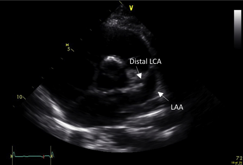
• LAA is a complex, variable anatomic structure that can be mistaken for other cardiac structures. • Color and spectral Doppler may help to discern the LAA from surrounding structures. • Cross-sectional imaging such as CTA can aid in defining the morphology of the LAA.

摘要

• 左心耳是一个复杂多变的解剖结构,可能会被误认为是其他心脏结构。

• 彩色及频谱多普勒有助于将左心耳与周围结构区分开来。

• 诸如CTA等横断面成像有助于明确左心耳的形态。

https://cdn.ncbi.nlm.nih.gov/pmc/blobs/9eee/8530799/ac8df45e15fa/gr5.jpg
https://cdn.ncbi.nlm.nih.gov/pmc/blobs/9eee/8530799/9e08467b54e8/fx1.jpg
https://cdn.ncbi.nlm.nih.gov/pmc/blobs/9eee/8530799/ee419f530210/gr1.jpg
https://cdn.ncbi.nlm.nih.gov/pmc/blobs/9eee/8530799/d2e7406cd307/gr2.jpg
https://cdn.ncbi.nlm.nih.gov/pmc/blobs/9eee/8530799/a967b669a7c5/gr3.jpg
https://cdn.ncbi.nlm.nih.gov/pmc/blobs/9eee/8530799/ef6a4380c633/gr4.jpg
https://cdn.ncbi.nlm.nih.gov/pmc/blobs/9eee/8530799/ac8df45e15fa/gr5.jpg

相似文献

1
A Case of a Left Atrial Appendage Disguised as a Coronary Artery Aneurysm.一例伪装成冠状动脉瘤的左心耳病例。
CASE (Phila). 2021 Aug 6;5(5):305-308. doi: 10.1016/j.case.2021.07.004. eCollection 2021 Oct.
2
Incremental benefit of three-dimensional transthoracic echocardiography in the assessment of left atrial appendage aneurysm leading to severe extrinsic compression of a coronary artery.三维经胸超声心动图在评估导致冠状动脉严重外在压迫的左心耳瘤中的增量效益。
Echocardiography. 2018 May;35(5):685-691. doi: 10.1111/echo.13901. Epub 2018 May 7.
3
Left atrial appendage aneurysm: A case report.左心耳动脉瘤:一例报告。
World J Clin Cases. 2020 Oct 6;8(19):4443-4449. doi: 10.12998/wjcc.v8.i19.4443.
4
Giant left atrial appendage aneurysm compressing the left anterior descending coronary artery.巨大左心耳动脉瘤压迫左前降支冠状动脉。
Echocardiography. 2016 Nov;33(11):1790-1792. doi: 10.1111/echo.13296. Epub 2016 Aug 19.
5
Congenital left atrial appendage aneurysm: Atypical presentation.先天性左心耳动脉瘤:非典型表现。
Ann Pediatr Cardiol. 2017 Sep-Dec;10(3):293-294. doi: 10.4103/apc.APC_4_17.
6
A Giant Left Circumflex Artery Aneurysm with Thrombosis Formation Presenting as a Left Atrioventricular Groove Mass.一例巨大左旋支动脉瘤伴血栓形成,表现为左房室沟肿块。
J Cardiovasc Echogr. 2021 Apr-Jun;31(2):116-118. doi: 10.4103/jcecho.jcecho_2_21. Epub 2021 Jul 28.
7
[Giant congenital intrapericardial left atrial appendage aneurysm: about a case and review of the literature].[巨大先天性心包内左心耳动脉瘤:一例报告并文献复习]
Pan Afr Med J. 2016 Jul 12;24:225. doi: 10.11604/pamj.2016.24.225.10012. eCollection 2016.
8
Late Presentation of Pulmonary Artery-Left Atrial Appendage Fistula Formation After Left Atrial Appendage Device Closure.左心耳封堵术后肺动脉-左心耳瘘形成的延迟表现
JACC Case Rep. 2020 May 20;2(5):814-818. doi: 10.1016/j.jaccas.2020.02.025. eCollection 2020 May.
9
Giant left atrial appendage with a common ventricular-appendicular wall and an abnormal course of the circumflex coronary artery in an asymptomatic 18-month-old girl.一名18个月大无症状女童,存在巨大左心耳,伴有共同的心室-心耳壁及回旋支冠状动脉走行异常。
Pediatr Cardiol. 2008 Mar;29(2):431-3. doi: 10.1007/s00246-007-9108-9. Epub 2007 Sep 26.
10
Giant congenital left atrial appendage aneurysm.巨大先天性左心耳动脉瘤
Cardiol Young. 2017 Apr;27(3):577-579. doi: 10.1017/S1047951116002791. Epub 2017 Jan 16.

引用本文的文献

1
Giant Left Atrial Appendage Aneurysm in a 6-Year-Old Girl with a Prothrombotic Genetic Predisposition: A Case Report and Literature Review.一名6岁患有血栓形成遗传易感性的女孩的巨大左心耳动脉瘤:病例报告及文献综述
Diagnostics (Basel). 2025 Aug 18;15(16):2070. doi: 10.3390/diagnostics15162070.
2
Left atrial appendage aneurysm: a descriptive systematic review of 177 cases.左心耳动脉瘤:177 例描述性系统评价。
BMC Cardiovasc Disord. 2024 Nov 11;24(1):633. doi: 10.1186/s12872-024-04323-x.

本文引用的文献

1
The left atrial appendage: anatomy, function, and noninvasive evaluation.左心耳:解剖、功能和无创评估。
JACC Cardiovasc Imaging. 2014 Dec;7(12):1251-65. doi: 10.1016/j.jcmg.2014.08.009.
2
Left atrial appendage: variations in morphology and position causing pitfalls in pediatric echocardiographic diagnosis.左心耳:形态和位置的变异导致小儿超声心动图诊断中的陷阱。
J Am Soc Echocardiogr. 2007 Aug;20(8):1011-6. doi: 10.1016/j.echo.2007.03.017. Epub 2007 Jun 25.
3
Echocardiographic assessment of the left atrial appendage.左心耳的超声心动图评估。
J Am Coll Cardiol. 1999 Dec;34(7):1867-77. doi: 10.1016/s0735-1097(99)00472-6.
4
Transthoracic Doppler echocardiography of normally originating coronary arteries in children.儿童正常起源冠状动脉的经胸多普勒超声心动图检查
J Am Soc Echocardiogr. 1998 May;11(5):409-20. doi: 10.1016/s0894-7317(98)70019-7.
5
Anatomy of the normal left atrial appendage: a quantitative study of age-related changes in 500 autopsy hearts: implications for echocardiographic examination.
Circulation. 1997 Nov 4;96(9):3112-5. doi: 10.1161/01.cir.96.9.3112.