Rodriguez-Falces Javier, Place Nicolas
Department of Electrical and Electronical Engineering, Public University of Navarre, Pamplona, Spain.
Institute of Sport Sciences, University of Lausanne, Lausanne, Switzerland.
Front Physiol. 2021 Oct 15;12:732624. doi: 10.3389/fphys.2021.732624. eCollection 2021.
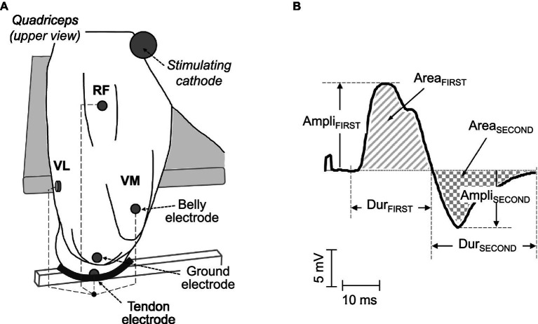
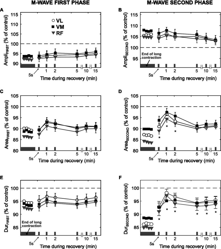
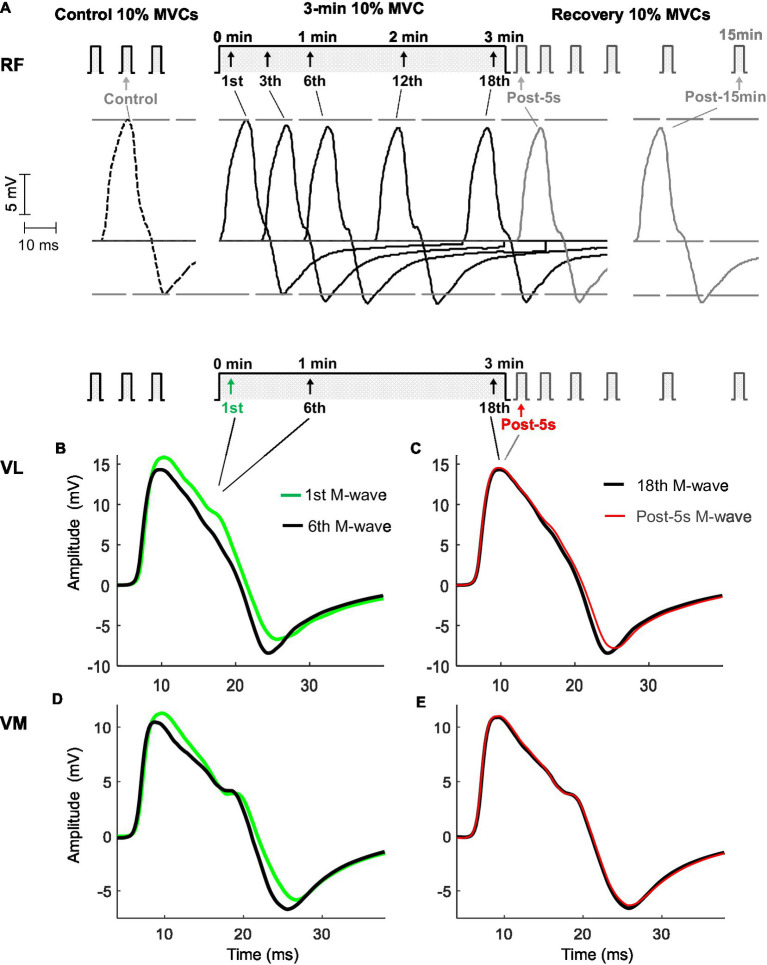
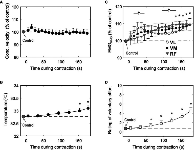
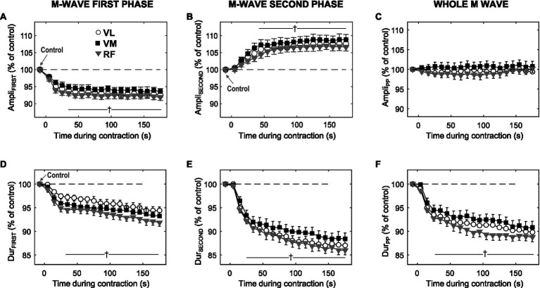
This study was undertaken to investigate whether sarcolemmal excitability is impaired during a sustained low-force contraction [10% maximal voluntary contraction (MVC)] by assessing muscle conduction velocity and also by analyzing separately the first and second phases of the muscle compound action potential (M wave). Twenty-one participants sustained an isometric knee extension of 10% MVC for 3min. M waves were evoked by supramaximal single shocks to the femoral nerve given at 10-s intervals. The amplitude, duration, and area of the first and second M-wave phases were computed. Muscle fiber conduction velocity, voluntary surface electromyographic (EMG), perceived effort, MVC force, peak twitch force, and temperature were also recorded. The main findings were: (1) During the sustained contraction, conduction velocity remained unchanged. (2) The amplitude of the M-wave first phase decreased for the first ~30s (-7%, <0.05) and stabilized thereafter, whereas the second phase amplitude increased for the initial ~30s (+7%, <0.05), before stabilizing. (3) Both duration and area decreased steeply during the first ~30s, and then more gradually for the rest of the contraction. (4) During the sustained contraction, perceived effort increased fivefold, whereas knee extension EMG increased by ~10%. (5) Maximal voluntary force and peak twitch force decreased (respectively, -9% and -10%, <0.05) after the low-force contraction. Collectively, the present results indicate that sarcolemmal excitability is well preserved during a sustained 10% MVC task. A depression of the M-wave first phase during a low-force contraction can occur even in the absence of changes in membrane excitability. The development of fatigue during a low-force contraction can occur without alteration of membrane excitability.
本研究旨在通过评估肌肉传导速度以及分别分析肌肉复合动作电位(M波)的第一和第二阶段,来探究在持续低强度收缩[10%最大自主收缩(MVC)]过程中肌膜兴奋性是否受损。21名参与者以10%MVC的强度持续进行等长伸膝动作3分钟。每隔10秒对股神经进行一次超强单刺激以诱发M波。计算第一和第二M波阶段的幅度、持续时间和面积。还记录了肌纤维传导速度、表面自愿肌电图(EMG)、主观用力感觉、MVC力量、峰值抽搐力量和温度。主要研究结果如下:(1)在持续收缩过程中,传导速度保持不变。(2)M波第一阶段的幅度在最初约30秒内下降(-7%,<0.05),此后稳定,而第二阶段的幅度在最初约30秒内增加(+7%,<0.05),之后稳定。(3)在最初约30秒内,持续时间和面积均急剧下降,在收缩的其余时间下降更为缓慢。(4)在持续收缩过程中,主观用力感觉增加了五倍,而伸膝EMG增加了约10%。(5)低强度收缩后,最大自主力量和峰值抽搐力量下降(分别为-9%和-10%,<0.05)。总体而言,目前的结果表明,在持续10%MVC任务过程中,肌膜兴奋性得到了很好的保留。即使在膜兴奋性没有变化的情况下,低强度收缩期间M波第一阶段也可能出现下降。低强度收缩期间疲劳的发展可能在膜兴奋性未改变的情况下发生。