Surboyo Meircurius Dwi Condro, Santosh Arvind Babu Rajendra, Hariyani Ninuk, Ernawati Diah Savitri, Cecilia Pamela Handy
Department of Oral Medicine, Faculty of Dental Medicine, Universitas Airlangga, Surabaya, 60132, Indonesia.
School of Dentistry, Faculty of Medical Sciences, University of the West Indies, Jamaica.
J Oral Biol Craniofac Res. 2021 Oct-Dec;11(4):618-623. doi: 10.1016/j.jobcr.2021.09.008. Epub 2021 Sep 16.
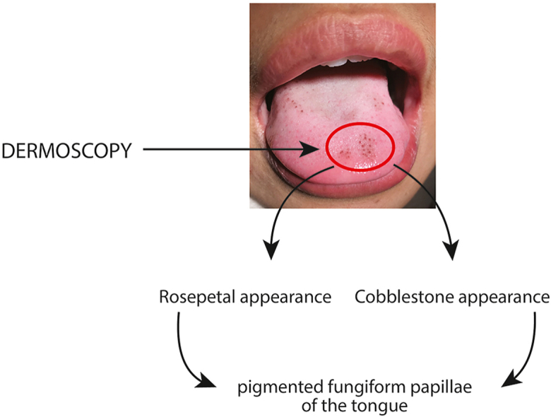
The practice of dermoscopy in dental and oral examination is low due to less popularity and not well established of the diagnostic tool in dental practice. The dermoscopy examination provides a specific dermoscopes details for pigmented papillary fungiform of tongue (PPFT) as cobblestone appearance and rose-petal appearance. With this dermoscopes details serves as a non-invasive diagnostic tool and prevents biopsy procedure.
We performed a systematic review to evaluate the published papers related to pigmented papillary fungiform on the tongue, aiming to understand the diagnostic role of dermoscopy examination in pigmented papillary fungiform.
Initial result was 136 studies. Final exclusion of 27 articles was made based on the following factors: reports with no clinical images, studies that did not confirm the diagnosis of PPFT and studies that did not use the dermoscopes details. Finally, seventeen studies with nineteen cases, reported of pigmented papillary fungiform of the tongue. Six studies (consist six cases) reported the dermoscopy and histopathology diagnosis of pigmented papillary fungiform, eleven studies (consist thirteen cases) reported only the dermoscopy. The dermoscopy examination presented cobblestone appearance is 47.37% and rose petal appearance is 52.63%. The comparation study by histopathology diagnosis was done, revealed no specific appearances.
The clinical appearance and dermoscopy is the key for diagnosis of the papillary fungiform on the tongue. Further research is needed for determining the etiology and predisposing factor in papillary fungiform so that the possibility of developing this condition can be predicted and proper treatment could be performed.
由于牙科学领域对皮肤镜检查的认知度较低且该诊断工具在牙科实践中尚未得到充分确立,因此其在口腔检查中的应用较少。皮肤镜检查为舌部色素性乳头状菌状瘤(PPFT)提供了特定的皮肤镜特征,如鹅卵石样外观和玫瑰花瓣样外观。凭借这些皮肤镜特征,其可作为一种非侵入性诊断工具,避免活检操作。
我们进行了一项系统综述,以评估与舌部色素性乳头状菌状瘤相关的已发表论文,旨在了解皮肤镜检查在色素性乳头状菌状瘤诊断中的作用。
初步检索结果为136项研究。基于以下因素最终排除了27篇文章:无临床图像的报告、未确诊为PPFT的研究以及未使用皮肤镜特征的研究。最终,有17项研究报告了19例舌部色素性乳头状菌状瘤病例。6项研究(包含6例病例)报告了色素性乳头状菌状瘤的皮肤镜和组织病理学诊断,11项研究(包含13例病例)仅报告了皮肤镜检查结果。皮肤镜检查呈现鹅卵石样外观的占47.37%,呈现玫瑰花瓣样外观的占52.63%。通过组织病理学诊断进行的对比研究显示无特异性表现。
临床表现和皮肤镜检查是诊断舌部乳头状菌状瘤的关键。需要进一步研究以确定乳头状菌状瘤的病因和易感因素,从而能够预测该疾病的发生可能性并进行恰当治疗。