Department of Oral & Maxillofacial Surgery, University of Groningen, University Medical Center Groningen, PO Box 30.001, 9700 RB, Groningen, the Netherlands.
Department of Pathology & Medical Biology, University of Groningen, University Medical Center Groningen, Groningen, the Netherlands.
Eur J Nucl Med Mol Imaging. 2022 Apr;49(5):1640-1649. doi: 10.1007/s00259-021-05567-x. Epub 2021 Nov 5.
Local recurrence occurs in ~ 19% of sinonasal inverted papilloma (SNIP) surgeries and is strongly associated with incomplete resection. During surgery, it is technically challenging to visualize and resect all SNIP tissue in this anatomically complex area. Proteins that are overexpressed in SNIP, such as vascular endothelial growth factor (VEGF), may serve as a target for fluorescence molecular imaging to guide surgical removal of SNIP. A proof-of-concept study was performed to investigate if the VEGF-targeted near-infrared fluorescent tracer bevacizumab-800CW specifically localizes in SNIP and whether it could be used as a clinical tool to guide SNIP surgery.
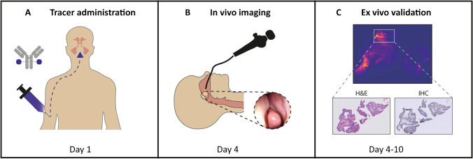
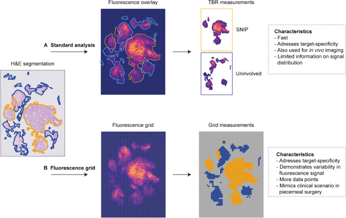
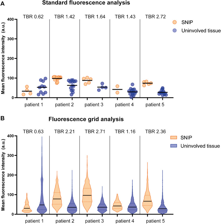
In five patients diagnosed with SNIP, 10 mg of bevacizumab-800CW was intravenously administered 3 days prior to surgery. Fluorescence molecular imaging was performed in vivo during surgery and ex vivo during the processing of the surgical specimen. Fluorescence signals were correlated with final histopathology and VEGF-A immunohistochemistry. We introduced a fluorescence grid analysis to assess the fluorescence signal in individual tissue fragments, due to the nature of the surgical procedure (i.e., piecemeal resection) allowing the detection of small SNIP residues and location of the tracer ex vivo.
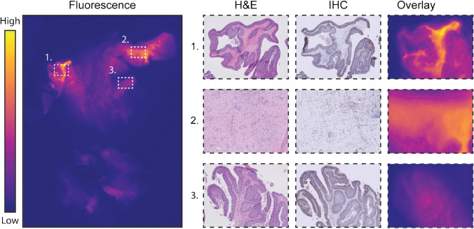
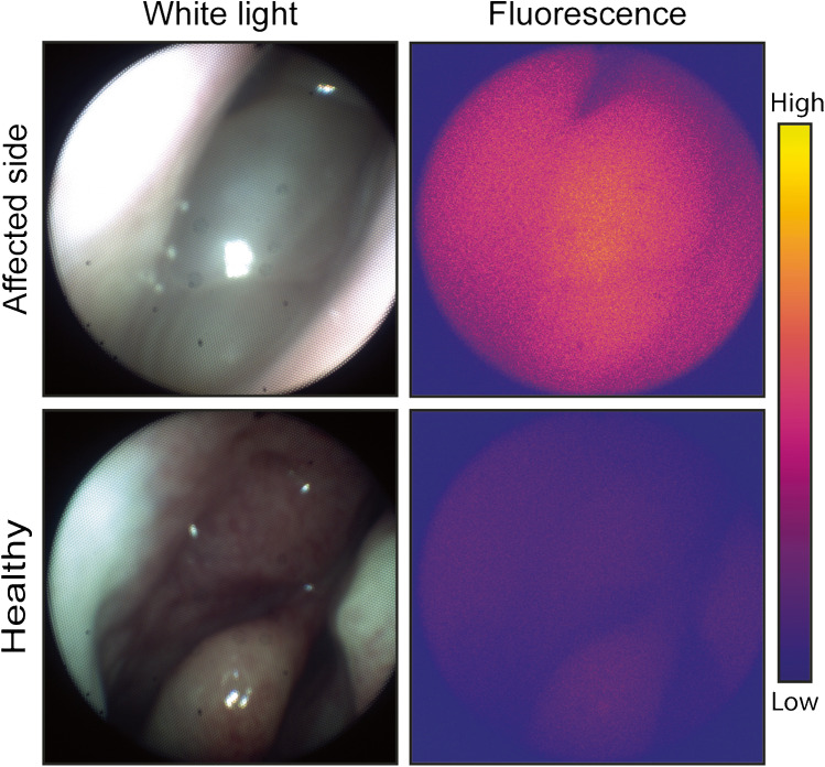
In all patients, fluorescence signal was detected in vivo during endoscopic SNIP surgery. Using ex vivo fluorescence grid analysis, we were able to correlate bevacizumab-800CW fluorescence of individual tissue fragments with final histopathology. Fluorescence grid analysis showed substantial variability in mean fluorescence intensity (FI), with SNIP tissue showing a median FI of 77.54 (IQR 50.47-112.30) compared to 35.99 (IQR 21.48-57.81) in uninvolved tissue (p < 0.0001), although the diagnostic ability was limited with an area under the curve of 0.78.
A fluorescence grid analysis could serve as a valid method to evaluate fluorescence molecular imaging in piecemeal surgeries. As such, although substantial differences were observed in fluorescence intensities, VEGF-A may not be the ideal target for SNIP surgery.
NCT03925285.
鼻内翻性乳头状瘤(SNIP)手术后约有 19%会发生局部复发,且与不完全切除密切相关。由于该解剖区域结构复杂,术中对 SNIP 组织进行可视化和切除具有一定难度。在 SNIP 中过度表达的蛋白,如血管内皮生长因子(VEGF),可能成为荧光分子成像的靶点,以指导 SNIP 的手术切除。本研究旨在探讨靶向 VEGF 的近红外荧光示踪剂贝伐单抗-800CW 是否能特异性定位于 SNIP,并能否作为一种临床工具来指导 SNIP 手术。
对 5 例 SNIP 患者,在术前 3 天静脉注射 10mg 贝伐单抗-800CW。术中进行体内荧光分子成像,手术标本处理过程中进行离体荧光分子成像。荧光信号与最终组织病理学和 VEGF-A 免疫组化结果相关联。由于手术过程为分片切除,为了评估每个组织碎片的荧光信号,我们引入了荧光网格分析,这种方法能够检测到小的 SNIP 残留并在离体时定位示踪剂。
所有患者在 SNIP 内镜手术中均能检测到体内荧光信号。通过离体荧光网格分析,我们能够将贝伐单抗-800CW 荧光与组织学最终结果进行关联。荧光网格分析显示,平均荧光强度(FI)存在显著差异,SNIP 组织的中位数为 77.54(IQR 50.47-112.30),而未受累组织的中位数为 35.99(IQR 21.48-57.81)(p<0.0001),但由于曲线下面积仅为 0.78,诊断能力有限。
荧光网格分析可以作为评估分片手术中荧光分子成像的有效方法。因此,尽管荧光强度存在显著差异,但 VEGF-A 可能不是 SNIP 手术的理想靶点。
NCT03925285。