Radiochemistry and Nuclear Imaging Group, CIC biomaGUNE, Basque Research and Technology Alliance (BRTA), 20014 San Sebastian, Spain.
Department of Nanomedicine and Theranostics, Institute for Experimental Molecular Imaging (ExMI), RWTH Aachen University Clinic and Helmholtz Institute for Biomedical Engineering, 52074 Aachen, Germany.
ACS Nano. 2021 Nov 23;15(11):16974-16981. doi: 10.1021/acsnano.1c09139. Epub 2021 Nov 8.
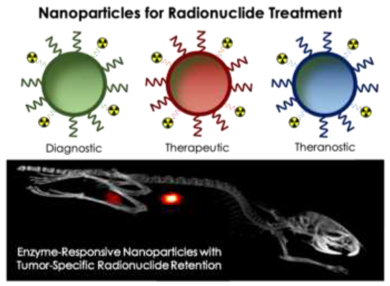
Nanoparticles have unique properties that can be exploited for cancer diagnosis and therapy. Intravenously injected nanoparticles accumulate predominantly in organs of the mononuclear phagocytic system, in addition to localizing in tumors and at sites of inflammation and infection. Accumulation in the liver and spleen lowers nanoparticles' ability to target pathological sites and compromises their use for radionuclide therapy. As described by Lee . in this issue of , radionuclide retention in liver and spleen can be greatly reduced by using liposomes that are surface-modified with esterase-cleavable radionuclide anchors. Because esterase activity is high in healthy tissues and low in tumors, the authors found that liposome-associated radioactivity rapidly cleared from the body and remained high only in tumors. The resulting images had high contrast-to-background ratios and remarkable tumor delineation. In this Perspective, we discuss these advances from early detection, cancer diagnosis, radionuclide therapy, and theranostics points of view. We outline the current clinical landscape of radionuclide targeting, imaging and therapy, and reflect on the roles that nanoparticles can play in these applications. We highlight the potential of nanoparticles that are responsive to endogenous stimuli for intraoperative imaging and, particularly, for individualized and improved radionuclide treatment. Taking these advances into account, future studies exploring the robustness and the clinical feasibility of nanomedicine-based radiotheranostic probes are eagerly awaited.
纳米粒子具有独特的性质,可用于癌症的诊断和治疗。静脉注射的纳米粒子主要积聚在单核吞噬细胞系统的器官中,除了在肿瘤和炎症及感染部位定位外。在肝脏和脾脏中的积聚降低了纳米粒子靶向病理部位的能力,并影响了它们在放射性核素治疗中的应用。正如 Lee 在本期杂志中所描述的,通过使用表面修饰有可被酯酶切割的放射性核素锚的脂质体,可以大大减少放射性核素在肝脏和脾脏中的滞留。由于酯酶活性在健康组织中较高,在肿瘤中较低,作者发现与脂质体相关的放射性物质迅速从体内清除,仅在肿瘤中保持高浓度。所得图像具有高对比度与背景比,并能显著描绘肿瘤。在本观点中,我们从早期检测、癌症诊断、放射性核素治疗和治疗学的角度讨论了这些进展。我们概述了放射性核素靶向、成像和治疗的当前临床状况,并思考了纳米粒子在这些应用中的作用。我们强调了对内源性刺激有反应的纳米粒子在术中成像,特别是在个体化和改进放射性核素治疗方面的潜力。考虑到这些进展,未来研究迫切需要探索基于纳米医学的放射治疗探针的稳健性和临床可行性。