• 文献检索
  • 文档翻译
  • 深度研究
  • 学术资讯
  • Suppr Zotero 插件Zotero 插件
  • 邀请有礼
  • 套餐&价格
  • 历史记录
应用&插件
Suppr Zotero 插件Zotero 插件浏览器插件Mac 客户端Windows 客户端微信小程序
定价
高级版会员购买积分包购买API积分包
服务
文献检索文档翻译深度研究API 文档MCP 服务
关于我们
关于 Suppr公司介绍联系我们用户协议隐私条款
关注我们

Suppr 超能文献

核心技术专利:CN118964589B侵权必究
粤ICP备2023148730 号-1Suppr @ 2026

文献检索

告别复杂PubMed语法,用中文像聊天一样搜索,搜遍4000万医学文献。AI智能推荐,让科研检索更轻松。

立即免费搜索

文件翻译

保留排版,准确专业,支持PDF/Word/PPT等文件格式,支持 12+语言互译。

免费翻译文档

深度研究

AI帮你快速写综述,25分钟生成高质量综述,智能提取关键信息,辅助科研写作。

立即免费体验

α-突触核蛋白在带负电荷的脂质囊泡上的取向:线性二色性揭示了螺旋结合模式的时间依赖性变化。

Orientation of α-Synuclein at Negatively Charged Lipid Vesicles: Linear Dichroism Reveals Time-Dependent Changes in Helix Binding Mode.

出版信息

J Am Chem Soc. 2021 Nov 17;143(45):18899-18906. doi: 10.1021/jacs.1c05344. Epub 2021 Nov 8.

DOI:10.1021/jacs.1c05344
PMID:34748321
原文链接:https://pmc.ncbi.nlm.nih.gov/articles/PMC8603351/
Abstract

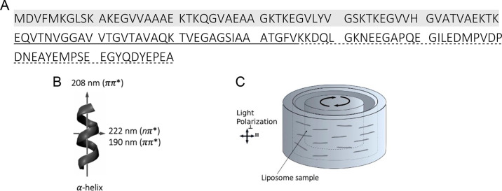
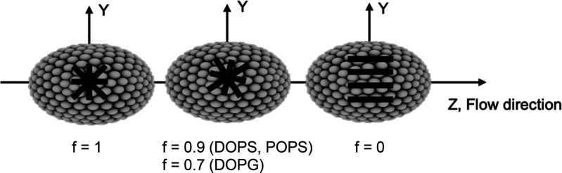
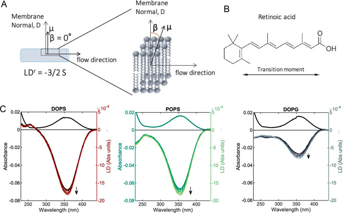
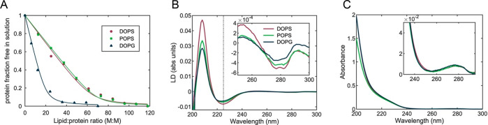
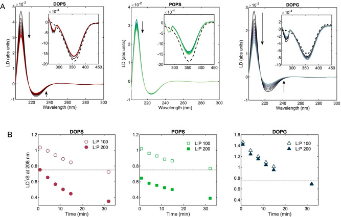
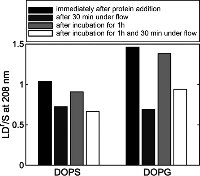
The neuronal protein α-synuclein, linked to Parkinson's disease, binds to negatively charged vesicles adopting a partial α-helix structure, but helix arrangement at the vesicle surface is not fully understood. Using linear dichroism spectroscopy (LD), we study the interaction of monomeric α-synuclein with large unilamellar vesicles of 1,2-dioleoyl--glycero-3-phospho-l-serine (DOPS), 1-palmitoyl-2-oleoyl--glycero-3-phospho-l-serine (POPS), and 1,2-dioleoyl--glycero-3-phospho-(1'--glycerol) (DOPG) under mild shear flow. The LD data of oriented lipid vesicles show that the long axis of the protein helix is oriented preferentially perpendicular to the membrane normal but deviates from a uniform in-plane distribution. Upon initial binding, a fraction of helices are oriented in the direction of least curvature for all ellipsoid-shaped vesicles at a lipid:protein molar ratio of 100. However, at a lower protein concentration the helices distribute uniformly on DOPS and POPS vesicles. In all cases, the α-synuclein helices rearrange with time (minute time scale) in the shear flow and begin to tilt into the vesicle membrane. Faster reorientation kinetics in the presence of flow suggests that modulation of membrane dynamics, by thermal or shear-dynamic activation, may overcome steric barriers by what may be called "flow catalysis".

摘要

神经元蛋白α-突触核蛋白与帕金森病有关,它与带负电荷的囊泡结合,形成部分α-螺旋结构,但囊泡表面的螺旋排列方式尚不完全清楚。我们使用线二色性光谱(LD)研究单体α-突触核蛋白与 1,2-二油酰基-sn-甘油-3-磷酸-l-丝氨酸(DOPS)、1-棕榈酰基-2-油酰基-sn-甘油-3-磷酸-l-丝氨酸(POPS)和 1,2-二油酰基-sn-甘油-3-磷酸-(1'-甘油)(DOPG)的大单室囊泡的相互作用,在温和的剪切流条件下进行。定向脂质囊泡的 LD 数据表明,蛋白质螺旋的长轴优先垂直于膜法线取向,但偏离了平面内的均匀分布。在初始结合时,对于所有各向异性囊泡,在脂质:蛋白质摩尔比为 100 的情况下,一部分螺旋沿最小曲率的方向取向。然而,在较低的蛋白质浓度下,DOPS 和 POPS 囊泡上的螺旋呈均匀分布。在所有情况下,α-突触核蛋白螺旋随时间(分钟时间尺度)在剪切流中重新排列,并开始倾斜进入囊泡膜。在存在流动的情况下更快的重取向动力学表明,通过热或剪切动力学激活来调节膜动力学,可能通过所谓的“流动催化”克服空间位阻。

https://cdn.ncbi.nlm.nih.gov/pmc/blobs/3c8d/8603351/e3e324e0bf1b/ja1c05344_0006.jpg
https://cdn.ncbi.nlm.nih.gov/pmc/blobs/3c8d/8603351/435742093117/ja1c05344_0001.jpg
https://cdn.ncbi.nlm.nih.gov/pmc/blobs/3c8d/8603351/5dc76d526c5d/ja1c05344_0002.jpg
https://cdn.ncbi.nlm.nih.gov/pmc/blobs/3c8d/8603351/9536b8f1651f/ja1c05344_0003.jpg
https://cdn.ncbi.nlm.nih.gov/pmc/blobs/3c8d/8603351/6b06b28a1c80/ja1c05344_0004.jpg
https://cdn.ncbi.nlm.nih.gov/pmc/blobs/3c8d/8603351/fda9ac1b0c68/ja1c05344_0005.jpg
https://cdn.ncbi.nlm.nih.gov/pmc/blobs/3c8d/8603351/e3e324e0bf1b/ja1c05344_0006.jpg

相似文献

1
Orientation of α-Synuclein at Negatively Charged Lipid Vesicles: Linear Dichroism Reveals Time-Dependent Changes in Helix Binding Mode.α-突触核蛋白在带负电荷的脂质囊泡上的取向:线性二色性揭示了螺旋结合模式的时间依赖性变化。
J Am Chem Soc. 2021 Nov 17;143(45):18899-18906. doi: 10.1021/jacs.1c05344. Epub 2021 Nov 8.
2
Discerning Dynamic Signatures of Membrane-Bound α-Synuclein Using Site-Specific Fluorescence Depolarization Kinetics.利用定点荧光各向异性动力学解析膜结合型 α-突触核蛋白的动态特征。
J Phys Chem B. 2020 Feb 6;124(5):708-717. doi: 10.1021/acs.jpcb.9b09118. Epub 2020 Jan 24.
3
Effect of lipid composition on the topography of membrane-associated hydrophobic helices: stabilization of transmembrane topography by anionic lipids.脂质组成对膜相关疏水螺旋结构的影响:阴离子脂质对跨膜结构的稳定作用。
J Mol Biol. 2008 Jun 13;379(4):704-18. doi: 10.1016/j.jmb.2008.04.026. Epub 2008 Apr 16.
4
Alpha-synuclein binds large unilamellar vesicles as an extended helix.α-突触核蛋白以伸展的螺旋形式结合大单层囊泡。
Biochemistry. 2009 Mar 24;48(11):2304-6. doi: 10.1021/bi900114z.
5
Evaluation of membrane models and their composition for islet amyloid polypeptide-membrane aggregation.用于胰岛淀粉样多肽-膜聚集的膜模型及其组成的评估。
Biochim Biophys Acta. 2013 Sep;1828(9):2091-8. doi: 10.1016/j.bbamem.2013.05.014. Epub 2013 May 23.
6
[The processes of α-synuclein amyloid protein complexes involved in the pathogenesis of Parkinson's disease].[参与帕金森病发病机制的α-突触核蛋白淀粉样蛋白复合物的过程]
Zh Nevrol Psikhiatr Im S S Korsakova. 2018;118(8):75-81. doi: 10.17116/jnevro201811808175.
7
α-Synuclein Oligomers Stabilize Pre-Existing Defects in Supported Bilayers and Propagate Membrane Damage in a Fractal-Like Pattern.α-突触核蛋白寡聚体稳定支持双层膜中预先存在的缺陷,并以类分形模式传播膜损伤。
Langmuir. 2016 Nov 15;32(45):11827-11836. doi: 10.1021/acs.langmuir.6b02572. Epub 2016 Nov 2.
8
Unraveling amyloid formation paths of Parkinson's disease protein α-synuclein triggered by anionic vesicles.解析帕金森病蛋白α-突触核蛋白在阴离子囊泡触发下的淀粉样形成途径。
Q Rev Biophys. 2017 Jan;50:e3. doi: 10.1017/S0033583517000026.
9
Intrinsic and membrane-facilitated α-synuclein oligomerization revealed by label-free detection through solid-state nanopores.通过固态纳米孔无标记检测揭示的内源性和膜促进的α-突触核蛋白寡聚化
Sci Rep. 2016 Feb 11;6:20776. doi: 10.1038/srep20776.
10
Two different binding modes of α-synuclein to lipid vesicles depending on its aggregation state.α-突触核蛋白根据其聚集状态与脂质囊泡的两种不同结合模式。
Biophys J. 2012 Apr 4;102(7):1646-55. doi: 10.1016/j.bpj.2012.01.059. Epub 2012 Apr 3.

引用本文的文献

1
Salt-Induced Membrane-Bound Conformation of the NAC Domain of α-Synuclein Leads to Structural Polymorphism of Amyloid Fibrils.盐诱导的α-突触核蛋白NAC结构域的膜结合构象导致淀粉样纤维的结构多态性。
Biomolecules. 2025 Mar 31;15(4):506. doi: 10.3390/biom15040506.
2
Order-to-Disorder and Disorder-to-Order Transitions of Proteins upon Binding to Phospholipid Membranes: Common Ground and Dissimilarities.蛋白质与磷脂膜结合时的有序-无序和无序-有序转变:共同点与差异
Biomolecules. 2025 Jan 30;15(2):198. doi: 10.3390/biom15020198.
3
Circular and Linear Dichroism for the Analysis of Small Noncoding RNA Properties.

本文引用的文献

1
Single-vesicle imaging reveals lipid-selective and stepwise membrane disruption by monomeric α-synuclein.单囊泡成像揭示单体α-突触核蛋白对脂质的选择性和逐步的膜破坏。
Proc Natl Acad Sci U S A. 2020 Jun 23;117(25):14178-14186. doi: 10.1073/pnas.1914670117. Epub 2020 Jun 8.
2
Avidity within the N-terminal anchor drives α-synuclein membrane interaction and insertion.N 端锚定结构域内的亲和力驱动α-突触核蛋白与膜相互作用和插入。
FASEB J. 2020 Jun;34(6):7462-7482. doi: 10.1096/fj.202000107R. Epub 2020 Apr 11.
3
Synaptic vesicle mimics affect the aggregation of wild-type and A53T α-synuclein variants differently albeit similar membrane affinity.
用于分析小非编码 RNA 性质的圆二色性和线二色性。
Methods Mol Biol. 2024;2741:399-416. doi: 10.1007/978-1-0716-3565-0_21.
4
Copper Binding and Redox Activity of α-Synuclein in Membrane-Like Environment.α-突触核蛋白在类似膜环境中的铜结合和氧化还原活性。
Biomolecules. 2023 Feb 3;13(2):287. doi: 10.3390/biom13020287.
5
The Consequences of GBA Deficiency in the Autophagy-Lysosome System in Parkinson's Disease Associated with GBA.帕金森病相关 GBA 中自噬溶酶体系统 GBA 缺乏的后果。
Cells. 2023 Jan 3;12(1):191. doi: 10.3390/cells12010191.
6
Association of Glial Activation and α-Synuclein Pathology in Parkinson's Disease.胶质细胞激活与帕金森病中α-突触核蛋白病理的关联。
Neurosci Bull. 2023 Mar;39(3):479-490. doi: 10.1007/s12264-022-00957-z. Epub 2022 Oct 14.
突触小泡模拟物尽管对膜亲和力相似,但对野生型和 A53T α-突触核蛋白变体的聚集有不同的影响。
Protein Eng Des Sel. 2019 Dec 13;32(2):59-66. doi: 10.1093/protein/gzz021.
4
Membrane-Protein-Hydration Interaction of α-Synuclein with Anionic Vesicles Probed via Angle-Resolved Second-Harmonic Scattering.通过角度分辨二次谐波散射研究α-突触核蛋白与阴离子囊泡的膜蛋白水合相互作用。
J Phys Chem B. 2019 Feb 7;123(5):1044-1049. doi: 10.1021/acs.jpcb.8b11096. Epub 2019 Jan 24.
5
Flow Alignment of Extracellular Vesicles: Structure and Orientation of Membrane-Associated Bio-macromolecules Studied with Polarized Light.细胞外囊泡的流型排列:用偏振光研究膜相关生物大分子的结构和取向。
Chembiochem. 2018 Mar 16;19(6):545-551. doi: 10.1002/cbic.201700378. Epub 2018 Feb 7.
6
Unraveling amyloid formation paths of Parkinson's disease protein α-synuclein triggered by anionic vesicles.解析帕金森病蛋白α-突触核蛋白在阴离子囊泡触发下的淀粉样形成途径。
Q Rev Biophys. 2017 Jan;50:e3. doi: 10.1017/S0033583517000026.
7
Parkinson disease.帕金森病。
Nat Rev Dis Primers. 2017 Mar 23;3:17013. doi: 10.1038/nrdp.2017.13.
8
Modulating membrane binding of α-synuclein as a therapeutic strategy.调节α-突触核蛋白的膜结合作为一种治疗策略。
Proc Natl Acad Sci U S A. 2017 Feb 7;114(6):1223-1225. doi: 10.1073/pnas.1620159114. Epub 2017 Jan 26.
9
Probing Microscopic Orientation in Membranes by Linear Dichroism.利用线性二色性探测膜中的微观取向
Langmuir. 2016 Mar 29;32(12):2841-6. doi: 10.1021/acs.langmuir.5b04229. Epub 2016 Mar 14.
10
Lipid vesicles trigger α-synuclein aggregation by stimulating primary nucleation.脂质囊泡通过刺激初级成核作用引发α-突触核蛋白聚集。
Nat Chem Biol. 2015 Mar;11(3):229-34. doi: 10.1038/nchembio.1750. Epub 2015 Feb 2.