Gupta Puneet, Jain Hitangee, Gill Misbah, Bharaj Gurpreet, Khalid Nauman, Chaudhry Waseem, Chhabra Lovely
Department of Interventional Cardiology, Northeast Ohio Medical University, Canton, OH 44272, United States.
BA-MD, Brooklyn College, Brooklyn, NY 11210, United States.
World J Cardiol. 2021 Oct 26;13(10):533-545. doi: 10.4330/wjc.v13.i10.533.
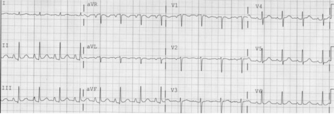
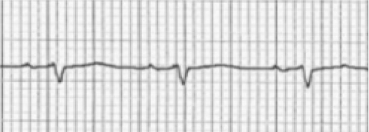
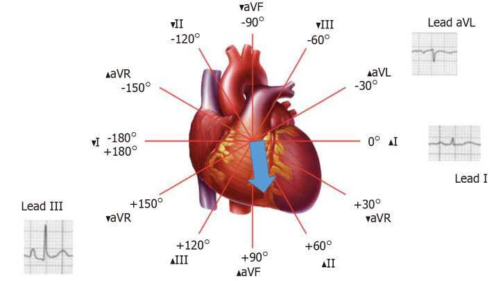
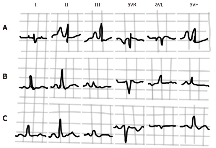
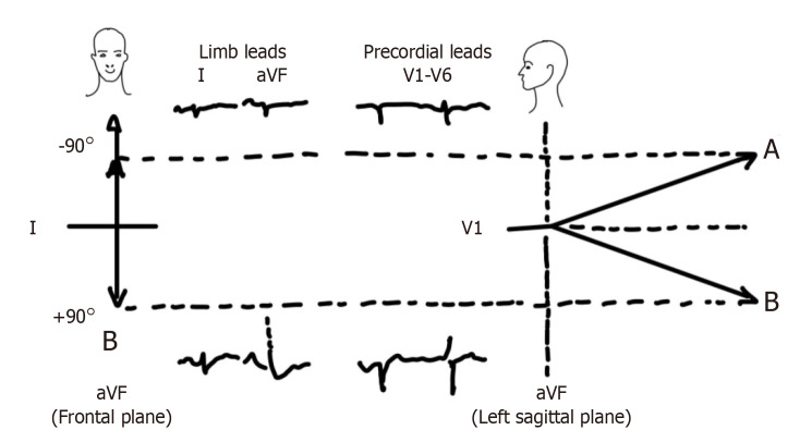
Chronic obstructive lung disease (COPD), predominantly emphysema, causes several thoracic anatomical and hemodynamic changes which may cause changes in various electrocardiographic parameters. A 12-lead electrocardiogram (ECG), which is often a part of routine evaluation in most clinical settings, may serve as a useful screening modality for diagnosis of COPD or emphysema. Our current article aims to provide a comprehensive review of the electrocardiographic changes encountered in COPD/emphysema utilizing published PubMed and Medline literature database. Several important ECG changes are present in COPD/emphysema and may serve as a good diagnostic tool. Verticalization of P-vector, changes in QRS duration, pattern recognition of precordial R-wave progression and axial shifts can be considered some of the most valuable markers among other changes. In conclusion, 12-lead surface electrocardiogram can serve as a valuable tool for the diagnosis of COPD and/or emphysema. An appropriate knowledge of these ECG changes can not only help in the diagnosis but can also immensely help in an appropriate clinical management of these patients.
慢性阻塞性肺疾病(COPD),主要是肺气肿,会引起多种胸部解剖学和血流动力学变化,这些变化可能导致各种心电图参数的改变。12导联心电图(ECG)在大多数临床环境中通常是常规评估的一部分,可作为诊断COPD或肺气肿的有用筛查手段。我们当前的文章旨在利用已发表的PubMed和Medline文献数据库,对COPD/肺气肿中出现的心电图变化进行全面综述。COPD/肺气肿中存在几种重要的心电图变化,可作为良好的诊断工具。P向量垂直化、QRS波时限变化、胸前导联R波进展的模式识别和电轴偏移可被视为其他变化中一些最有价值的标志物。总之,12导联体表心电图可作为诊断COPD和/或肺气肿的有价值工具。对这些心电图变化的适当了解不仅有助于诊断,还能极大地帮助对这些患者进行适当的临床管理。