• 文献检索
  • 文档翻译
  • 深度研究
  • 学术资讯
  • Suppr Zotero 插件Zotero 插件
  • 邀请有礼
  • 套餐&价格
  • 历史记录
应用&插件
Suppr Zotero 插件Zotero 插件浏览器插件Mac 客户端Windows 客户端微信小程序
定价
高级版会员购买积分包购买API积分包
服务
文献检索文档翻译深度研究API 文档MCP 服务
关于我们
关于 Suppr公司介绍联系我们用户协议隐私条款
关注我们

Suppr 超能文献

核心技术专利:CN118964589B侵权必究
粤ICP备2023148730 号-1Suppr @ 2026

文献检索

告别复杂PubMed语法,用中文像聊天一样搜索,搜遍4000万医学文献。AI智能推荐,让科研检索更轻松。

立即免费搜索

文件翻译

保留排版,准确专业,支持PDF/Word/PPT等文件格式,支持 12+语言互译。

免费翻译文档

深度研究

AI帮你快速写综述,25分钟生成高质量综述,智能提取关键信息,辅助科研写作。

立即免费体验

影像引导下经皮热消融治疗小的单发肝细胞癌的最佳消融边缘及基于消融边缘的局部复发预测:一项多中心研究

An Optimal Ablative Margin of Small Single Hepatocellular Carcinoma Treated with Image-Guided Percutaneous Thermal Ablation and Local Recurrence Prediction Base on the Ablative Margin: A Multicenter Study.

作者信息

Li Feng-Yao, Li Jian-Guo, Wu Song-Song, Ye Huo-Lin, He Xu-Qi, Zeng Qing-Jing, Zheng Rong-Qin, An Chao, Li Kai

机构信息

Department of Ultrasound, Guangdong Key Laboratory of Liver Disease Research, The Third Affiliated Hospital of Sun Yat-sen University, Guangzhou, People's Republic of China.

The Department of Infectious Disease,The Third Affiliated Hospital of Sun Yat-sen University, Guangzhou, People's Republic of China.

出版信息

J Hepatocell Carcinoma. 2021 Nov 15;8:1375-1388. doi: 10.2147/JHC.S330746. eCollection 2021.

DOI:10.2147/JHC.S330746
PMID:34815974
原文链接:https://pmc.ncbi.nlm.nih.gov/articles/PMC8604653/
Abstract

OBJECTIVE

To explore the best ablative margin (AM) for single hepatocellular carcinoma (HCC) patients with image-guided percutaneous thermal ablation (IPTA) based on MRI-MRI fusion imaging, and to develop and validate a local tumor progression (LTP) predictive model based on the recommended AM.

METHODS

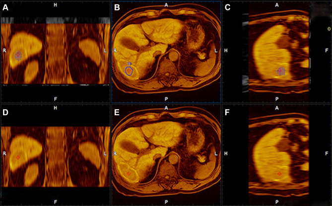
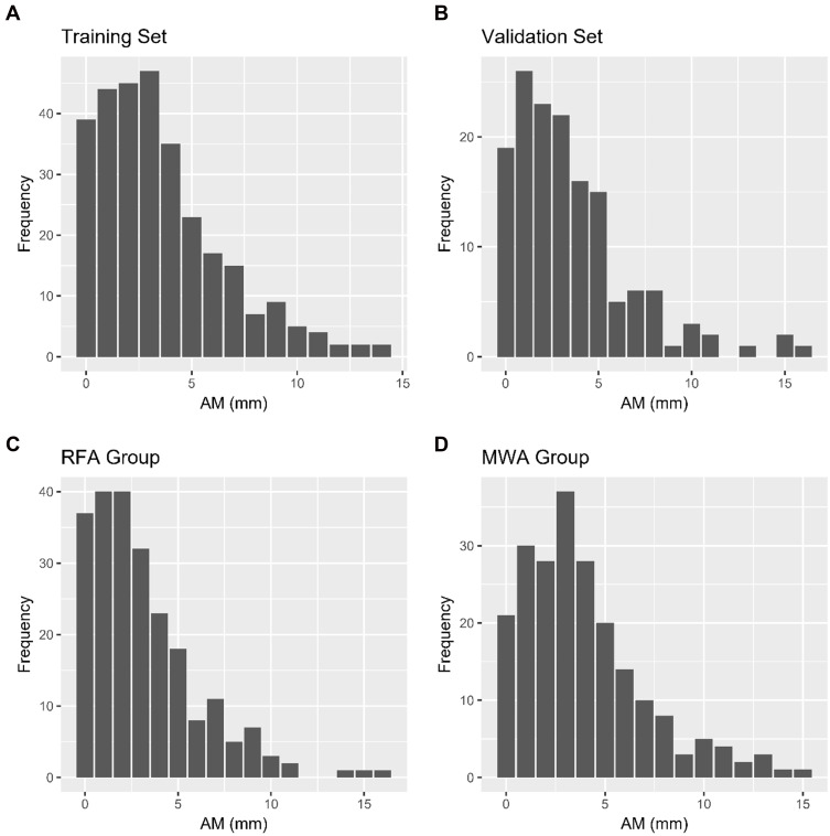
Between March 2014 and August 2019, 444 treatment-naïve patients with single HCC (diameter ≤3 cm) who underwent IPTA as first-line treatment from three hospitals were included, which were randomly divided into training (n= 296) and validation (n = 148) cohorts. We measured the ablative margin (AM) by MRI-MRI fusion imaging based on pre-ablation and post-ablation images. Then, we followed up their LPT and verified the optimal AM. Risk factors related to LTP were explored through Cox regression models, the nomogram was developed to predict the LTP risk base on the risk factors, and subsequently validated. The predictive performance and discrimination were assessed and compared with conventional indices.

RESULTS

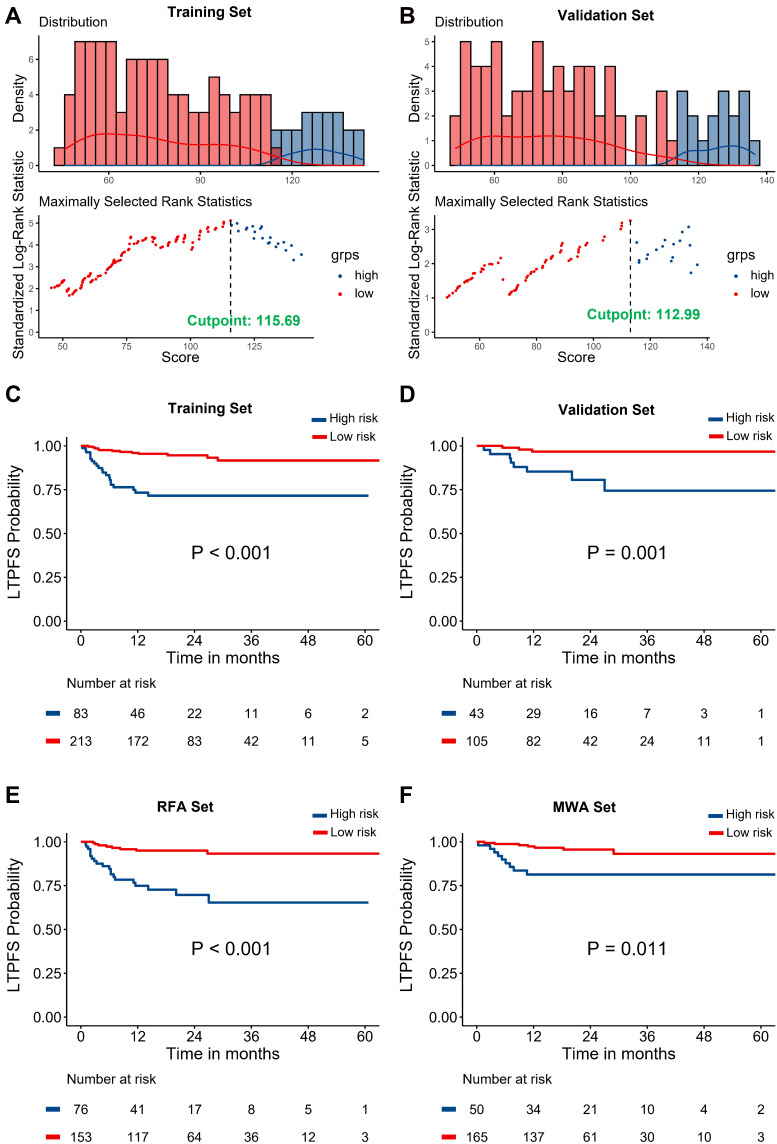
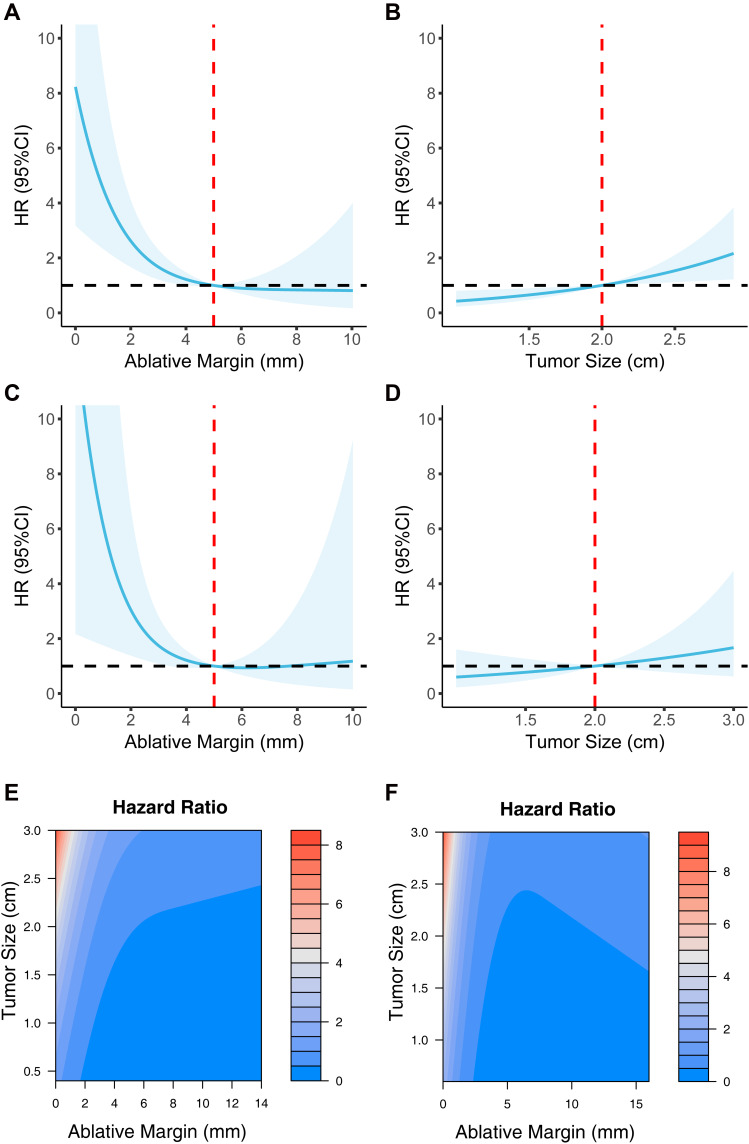
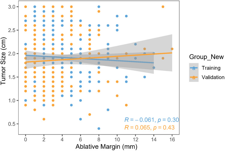
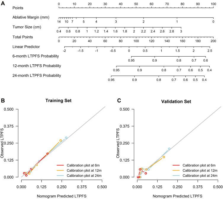
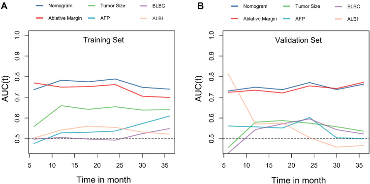
The median follow-up was 19.9 months (95% CI 18.0-21.8) for the entire cohort. The results revealed that the tumor size (HR: 2.16; 95% CI 1.25-3.72; P = 0.003) and AM (HR: 0.72; 95% CI, 0.61-0.85; P < 0.001) were independent prognostic factors for LTP. The AM had a pronounced nonlinear impact on LTP, and a cut-off value of 5-mm was optimal. We developed and validated an LTP predictive model based on the linear tumor size and nonlinear AM. The model showed good predictive accuracy and discrimination (training set, concordance index [C-index] of 0.751; validation set, C-index of 0.756) and outperformed other conventional indices.

CONCLUSION

The 5-mm AM is recommended for the best IPTA candidates with single HCC (diameter ≤3 cm). We provided an LTP predictive model that exhibited adequate performance for individualized prediction and risk stratification.

摘要

目的

基于磁共振成像-磁共振成像融合成像,探讨影像引导下经皮热消融(IPTA)治疗单发肝细胞癌(HCC)患者的最佳消融边缘(AM),并基于推荐的AM建立和验证局部肿瘤进展(LTP)预测模型。

方法

纳入2014年3月至2019年8月期间来自三家医院的444例未经治疗的单发HCC(直径≤3 cm)患者,这些患者接受IPTA作为一线治疗,并随机分为训练队列(n = 296)和验证队列(n = 148)。我们根据消融前和消融后的图像,通过磁共振成像-磁共振成像融合成像测量消融边缘(AM)。然后,我们对他们的LPT进行随访,并验证最佳AM。通过Cox回归模型探索与LTP相关的危险因素,基于危险因素建立列线图以预测LTP风险,随后进行验证。评估预测性能和辨别力,并与传统指标进行比较。

结果

整个队列的中位随访时间为19.9个月(95% CI 18.0 - 21.8)。结果显示,肿瘤大小(HR:2.16;95% CI 1.25 - 3.72;P = 0.003)和AM(HR:0.72;95% CI,0.61 - 0.85;P < 0.001)是LTP的独立预后因素。AM对LTP有明显的非线性影响,5 mm的截断值是最佳的。我们基于线性肿瘤大小和非线性AM建立并验证了一个LTP预测模型。该模型显示出良好的预测准确性和辨别力(训练集,一致性指数[C-index]为0.751;验证集,C-index为0.756),并且优于其他传统指标。

结论

对于最佳的单发HCC(直径≤3 cm)IPTA候选患者,建议采用5 mm的AM。我们提供了一个LTP预测模型,该模型在个体化预测和风险分层方面表现出足够的性能。

https://cdn.ncbi.nlm.nih.gov/pmc/blobs/ea12/8604653/2328462fa75d/JHC-8-1375-g0007.jpg
https://cdn.ncbi.nlm.nih.gov/pmc/blobs/ea12/8604653/905320da23a4/JHC-8-1375-g0001.jpg
https://cdn.ncbi.nlm.nih.gov/pmc/blobs/ea12/8604653/4ddb67530ca3/JHC-8-1375-g0002.jpg
https://cdn.ncbi.nlm.nih.gov/pmc/blobs/ea12/8604653/d471e44729c4/JHC-8-1375-g0003.jpg
https://cdn.ncbi.nlm.nih.gov/pmc/blobs/ea12/8604653/1542622197ab/JHC-8-1375-g0004.jpg
https://cdn.ncbi.nlm.nih.gov/pmc/blobs/ea12/8604653/20b16342713c/JHC-8-1375-g0005.jpg
https://cdn.ncbi.nlm.nih.gov/pmc/blobs/ea12/8604653/4c0acdf6b670/JHC-8-1375-g0006.jpg
https://cdn.ncbi.nlm.nih.gov/pmc/blobs/ea12/8604653/2328462fa75d/JHC-8-1375-g0007.jpg

相似文献

1
An Optimal Ablative Margin of Small Single Hepatocellular Carcinoma Treated with Image-Guided Percutaneous Thermal Ablation and Local Recurrence Prediction Base on the Ablative Margin: A Multicenter Study.影像引导下经皮热消融治疗小的单发肝细胞癌的最佳消融边缘及基于消融边缘的局部复发预测:一项多中心研究
J Hepatocell Carcinoma. 2021 Nov 15;8:1375-1388. doi: 10.2147/JHC.S330746. eCollection 2021.
2
Assessment of Ablative Margin After Microwave Ablation for Hepatocellular Carcinoma Using Deep Learning-Based Deformable Image Registration.基于深度学习的可变形图像配准技术在肝细胞癌微波消融术后消融边缘评估中的应用
Front Oncol. 2020 Sep 24;10:573316. doi: 10.3389/fonc.2020.573316. eCollection 2020.
3
Predictive Value of Ablative Margin Assessment After Microwave Ablation for Local Tumor Progression in Medium and Large Hepatocellular Carcinoma: Computed Tomography-Computed Tomography Image Fusion Method Versus Side-by-Side Method.微波消融治疗中大型肝癌后消融边界评估对局部肿瘤进展的预测价值:CT-CT 图像融合法与并排法比较。
J Comput Assist Tomogr. 2023;47(1):31-37. doi: 10.1097/RCT.0000000000001395. Epub 2022 Nov 4.
4
Prediction of Local Tumor Progression after Radiofrequency Ablation (RFA) of Hepatocellular Carcinoma by Assessment of Ablative Margin Using Pre-RFA MRI and Post-RFA CT Registration.基于术前 RFA MRI 与术后 RFA CT 配准评估消融边界预测肝癌射频消融后局部肿瘤进展
Korean J Radiol. 2018 Nov-Dec;19(6):1053-1065. doi: 10.3348/kjr.2018.19.6.1053. Epub 2018 Oct 18.
5
The minimal ablative margin of radiofrequency ablation of hepatocellular carcinoma (> 2 and < 5 cm) needed to prevent local tumor progression: 3D quantitative assessment using CT image fusion.射频消融治疗肝细胞癌(>2cm 且<5cm)所需的最小消融边界以预防局部肿瘤进展:使用 CT 图像融合的 3D 定量评估。
AJR Am J Roentgenol. 2010 Sep;195(3):758-65. doi: 10.2214/AJR.09.2954.
6
A novel nomogram to predict the local tumor progression after microwave ablation in patients with early-stage hepatocellular carcinoma: A tool in prediction of successful ablation.一种用于预测早期肝细胞癌患者微波消融后局部肿瘤进展的新诺莫图:一种预测消融成功的工具。
Cancer Med. 2020 Jan;9(1):104-115. doi: 10.1002/cam4.2606. Epub 2019 Nov 12.
7
The tumor ghost on MRI after microwave ablation for hepatocellular carcinoma: A new modality to assess the ablative margin.肝癌微波消融治疗后 MRI 上的肿瘤鬼影:评估消融边界的新方式。
Eur J Radiol. 2023 Jan;158:110617. doi: 10.1016/j.ejrad.2022.110617. Epub 2022 Nov 24.
8
Improvement of ablative margins by the intraoperative use of CEUS-CT/MR image fusion in hepatocellular carcinoma.在肝细胞癌中通过术中使用CEUS-CT/MR图像融合改善消融边缘
BMC Cancer. 2016 Apr 18;16:277. doi: 10.1186/s12885-016-2306-1.
9
Minimal ablative margin (MAM) assessment with image fusion: an independent predictor for local tumor progression in hepatocellular carcinoma after stereotactic radiofrequency ablation.采用图像融合技术评估最小消融边界(MAM):对立体定向射频消融后肝细胞癌局部肿瘤进展的独立预测因子。
Eur Radiol. 2020 May;30(5):2463-2472. doi: 10.1007/s00330-019-06609-7. Epub 2020 Jan 30.
10
Imaging evaluation of ablative margin and index tumor immediately after radiofrequency ablation for hepatocellular carcinoma: comparison between multidetector-row CT and MR imaging.多排螺旋 CT 与 MR 成像在肝癌射频消融术后即刻评估消融边界和肿瘤指数的比较。
Abdom Radiol (NY). 2017 Oct;42(10):2527-2537. doi: 10.1007/s00261-017-1146-z.

引用本文的文献

1
Clinical and Imaging-Based Prognostic Models for Recurrence and Local Tumor Progression Following Thermal Ablation of Hepatocellular Carcinoma: A Systematic Review.肝细胞癌热消融术后复发和局部肿瘤进展的基于临床和影像的预后模型:一项系统评价
Cancers (Basel). 2025 Aug 14;17(16):2656. doi: 10.3390/cancers17162656.
2
Liver transplantation for malignant liver tumors.恶性肝肿瘤的肝移植
ILIVER. 2022 Apr 18;1(1):3-11. doi: 10.1016/j.iliver.2022.04.002. eCollection 2022 Mar.
3
Can Intra-Operative Ablation-Specific Features Based on Ultrasound Fusion Imaging be Used to Predict Early Recurrence of Hepatocellular Carcinoma After Microwave Ablation: A Proof-of-Concept Study.

本文引用的文献

1
EASL clinical practice guidelines for the management of occupational liver diseases.欧洲肝脏研究学会职业性肝病管理临床实践指南
Liver Int. 2020 Feb;40 Suppl 1:136-141. doi: 10.1111/liv.14349.
2
Multimodal Percutaneous Thermal Ablation of Small Hepatocellular Carcinoma: Predictive Factors of Recurrence and Survival in Western Patients.小肝细胞癌的多模态经皮热消融:西方患者复发和生存的预测因素
Cancers (Basel). 2020 Jan 29;12(2):313. doi: 10.3390/cancers12020313.
3
Minimal ablative margin (MAM) assessment with image fusion: an independent predictor for local tumor progression in hepatocellular carcinoma after stereotactic radiofrequency ablation.
基于超声融合成像的术中消融特异性特征能否用于预测肝细胞癌微波消融术后早期复发:一项概念验证研究
J Hepatocell Carcinoma. 2025 May 12;12:949-960. doi: 10.2147/JHC.S512926. eCollection 2025.
4
Long-Term Analysis of Recurrence Beyond Milan Criteria Following Ablation of Solitary Early-Stage Hepatocellular Carcinoma ≤3 cm in Potentially Transplantable Patients: A Over 10-Year Survival Study.潜在可移植患者中直径≤3cm的孤立早期肝细胞癌消融术后超出米兰标准的复发长期分析:一项超过10年的生存研究
J Hepatocell Carcinoma. 2025 Feb 3;12:205-218. doi: 10.2147/JHC.S505979. eCollection 2025.
5
Precise liver tumor ablation: the clinical potential of US-US overlay fusion guidance.精准肝肿瘤消融:超声-超声叠加融合引导的临床潜力
Ultrasonography. 2024 Nov;43(6):407-412. doi: 10.14366/usg.24133. Epub 2024 Aug 13.
6
Identification of proliferative hepatocellular carcinoma using the SMARS score and implications for microwave ablation.使用SMARS评分识别增殖性肝细胞癌及其对微波消融的意义。
Insights Imaging. 2024 Sep 10;15(1):220. doi: 10.1186/s13244-024-01792-8.
7
3D-CEUS/MRI-CEUS fusion imaging vs 2D-CEUS after locoregional therapies for hepatocellular carcinoma: a multicenter prospective study of therapeutic response evaluation.肝细胞癌局部区域治疗后3D-CEUS/MRI-CEUS融合成像与二维CEUS对比:治疗反应评估的多中心前瞻性研究
Eur Radiol. 2025 Jan;35(1):453-462. doi: 10.1007/s00330-024-10915-0. Epub 2024 Jul 11.
8
Single-arm prospective study comparing ablation zone volume between time zero and 24 h after microwave ablation of liver tumors.一项单臂前瞻性研究,比较肝肿瘤微波消融术后零时与24小时之间的消融区体积。
Abdom Radiol (NY). 2024 Sep;49(9):3136-3142. doi: 10.1007/s00261-024-04185-z. Epub 2024 Feb 24.
9
Mechanisms and therapeutic strategies to combat the recurrence and progression of hepatocellular carcinoma after thermal ablation.热消融后肝细胞癌复发与进展的机制及治疗策略
J Interv Med. 2023 Oct 18;6(4):160-169. doi: 10.1016/j.jimed.2023.10.004. eCollection 2023 Nov.
10
Multiparametric MRI-based model for prediction of local progression of hepatocellular carcinoma after thermal ablation.基于多参数 MRI 的模型预测肝癌热消融治疗后局部进展。
Cancer Med. 2023 Sep;12(17):17529-17540. doi: 10.1002/cam4.6277. Epub 2023 Sep 11.
采用图像融合技术评估最小消融边界(MAM):对立体定向射频消融后肝细胞癌局部肿瘤进展的独立预测因子。
Eur Radiol. 2020 May;30(5):2463-2472. doi: 10.1007/s00330-019-06609-7. Epub 2020 Jan 30.
4
A phenotypical map of disseminated hepatocellular carcinoma suggests clonal constraints in metastatic sites.播散性肝细胞癌的表型图谱提示转移部位存在克隆限制。
Histopathology. 2019 Apr;74(5):718-730. doi: 10.1111/his.13809. Epub 2019 Feb 21.
5
Outcomes of radiofrequency ablation as first-line therapy for hepatocellular carcinoma less than 3 cm in potentially transplantable patients.射频消融作为潜在可移植患者小于 3cm 的肝细胞癌一线治疗的结果。
J Hepatol. 2019 May;70(5):866-873. doi: 10.1016/j.jhep.2018.12.027. Epub 2019 Jan 5.
6
Radiofrequency ablation versus surgical resection of hepatocellular carcinoma: contemporary treatment trends and outcomes from the United States National Cancer Database.射频消融与手术切除治疗肝细胞癌:来自美国国家癌症数据库的当代治疗趋势和结果。
Eur Radiol. 2019 May;29(5):2679-2689. doi: 10.1007/s00330-018-5902-4. Epub 2018 Dec 17.
7
Impact of Anatomical Resection for Hepatocellular Carcinoma With Microportal Invasion (vp1): A Multi-institutional Study by the Kyushu Study Group of Liver Surgery.解剖性肝切除术治疗微门静脉侵犯肝细胞癌(vp1)的影响:九州肝外科学会多机构研究。
Ann Surg. 2020 Feb;271(2):339-346. doi: 10.1097/SLA.0000000000002981.
8
No-touch multibipolar radiofrequency ablation vs. surgical resection for solitary hepatocellular carcinoma ranging from 2 to 5 cm.无接触式多点射频消融与手术切除治疗 2-5cm 单发肝细胞癌的比较。
J Hepatol. 2018 Jun;68(6):1172-1180. doi: 10.1016/j.jhep.2018.01.014. Epub 2018 Feb 2.
9
Safety margin after radiofrequency ablation of hepatocellular carcinoma: precise assessment with a three-dimensional reconstruction technique using CT imaging.肝癌射频消融术后安全边界:应用 CT 成像三维重建技术精确评估。
Int J Hyperthermia. 2018 Dec;34(8):1135-1141. doi: 10.1080/02656736.2017.1411981. Epub 2018 Feb 2.
10
Local recurrence after microwave thermosphere ablation of malignant liver tumors: results of a surgical series.微波热疗消融治疗肝脏恶性肿瘤后的局部复发:外科系列研究结果。
Surgery. 2018 Apr;163(4):709-713. doi: 10.1016/j.surg.2017.10.026. Epub 2017 Dec 19.