Gomez Cifuentes Juan David, Haider Mahnur, Sanaka Madhusudhan R, Kumar Prabhat, Bena James, McMichael John, Sohal Davendra P, Raja Siva, Murthy Sudish, Thota Prashanthi N
Department of Gastroenterology and Hepatology, Baylor College of Medicine, Houston, USA.
Section of General Internal Medicine, Tulane Medical Center, New Orleans, USA.
Cureus. 2021 Oct 23;13(10):e18991. doi: 10.7759/cureus.18991. eCollection 2021 Oct.
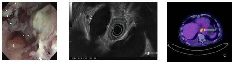
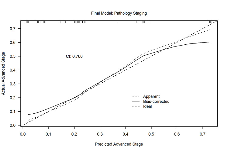
Background In patients with resectable esophageal adenocarcinoma (EAC), the decision for neoadjuvant treatment depends on clinical staging with endoscopic ultrasound (EUS) and positron-emission tomography (PET) scan. Patients with locally advanced EAC pathology misclassified as early EAC by clinical staging are missing the opportunity to receive neoadjuvant therapy. We aim to identify predictors of locally advanced pathology in EAC to determine more accurately those who benefit from neoadjuvant therapy. Methods Retrospective study of patients who underwent upfront endoscopic or surgical resection for EAC without neoadjuvant therapy from January 2011 to December 2017 was performed. Clinical characteristics, EUS, PET scan and histologic findings were analyzed. Multivariable analysis of predictors of locally advanced stage was performed and a risk prediction score was developed. Results A total of 97 patients were included; 68 patients were staged as early EAC (pT1 or pT2 and pN0) and 29 patients were staged as locally advanced EAC (pT1 or pT2 with pN1 and pT3 or pT4 irrespective of N status). In a predictive model of EAC, patients presenting with dysphagia, tumor size >2 cm, exophytic mass appearance on endoscopy and absence of hiatal hernia were more likely to be have locally advanced pathology with a probability of 70% (C-statistic 0.766). Conclusions A risk prediction model based on the presence of dysphagia, tumor size >2 cm, exophytic mass appearance and absence of hiatal hernia can be used to identify locally advanced pathology in EAC patients.
背景 在可切除食管腺癌(EAC)患者中,新辅助治疗的决策取决于内镜超声(EUS)和正电子发射断层扫描(PET)扫描的临床分期。临床分期将局部晚期EAC病理误诊为早期EAC的患者失去了接受新辅助治疗的机会。我们旨在确定EAC中局部晚期病理的预测因素,以更准确地确定那些能从新辅助治疗中获益的患者。
方法 对2011年1月至2017年12月期间未经新辅助治疗直接接受内镜或手术切除的EAC患者进行回顾性研究。分析临床特征、EUS、PET扫描和组织学结果。对局部晚期阶段的预测因素进行多变量分析,并制定风险预测评分。
结果 共纳入97例患者;68例患者分期为早期EAC(pT1或pT2且pN0),29例患者分期为局部晚期EAC(pT1或pT2伴pN1以及pT3或pT4,不考虑N状态)。在EAC预测模型中,出现吞咽困难、肿瘤大小>2 cm、内镜下外生性肿块外观以及无食管裂孔疝的患者更有可能患有局部晚期病理,概率为70%(C统计量0.766)。
结论 基于吞咽困难、肿瘤大小>2 cm、外生性肿块外观和无食管裂孔疝的风险预测模型可用于识别EAC患者的局部晚期病理。