Laboratory of Cerebrovascular Research, Montreal Neurological Institute, McGill University, Montreal, Quebec, Canada.
Br J Pharmacol. 2022 May;179(10):2259-2274. doi: 10.1111/bph.15751. Epub 2022 Feb 10.
Inward rectifier potassium (K ) channels are key effectors of vasodilatation in neurovascular coupling (NVC). K channels expressed in cerebral endothelial cells (ECs) have been confirmed as essential modulators of NVC. Alzheimer's disease (AD) and cerebrovascular disease (CVD) impact on EC-K channel function, but whether oxidative stress or inflammation explains this impairment remains elusive.
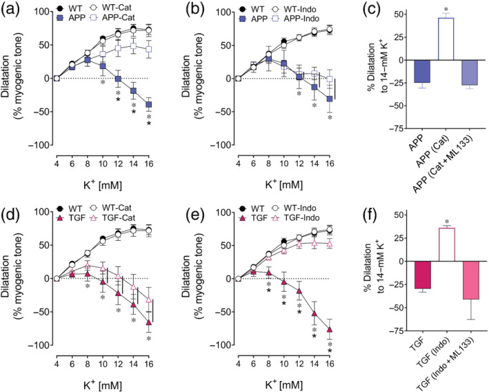
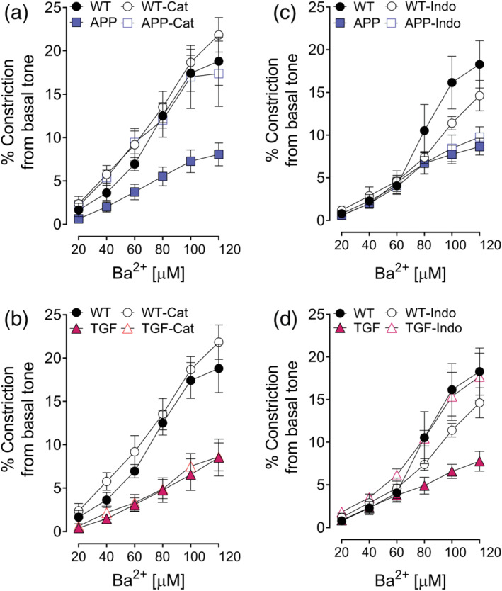
We evaluated K channel function in intact and EC-denuded pial arteries of wild-type (WT) and transgenic mice overexpressing a mutated form of the human amyloid precursor protein (APP mice, recapitulating amyloid β-induced oxidative stress seen in AD) or a constitutively active form of TGF-β1 (TGF mice, recapitulating inflammation seen in cerebrovascular pathology). The benefits of antioxidant (catalase) or anti-inflammatory (indomethacin) drugs also were investigated. Vascular and neuronal components of NVC were assessed in vivo.
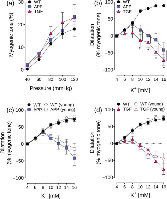
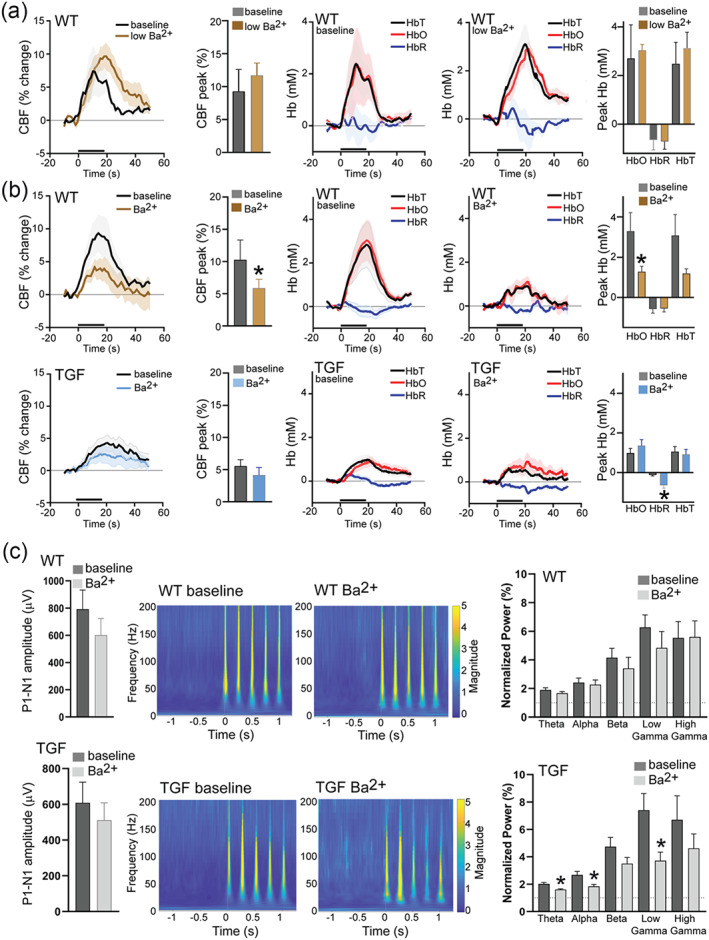
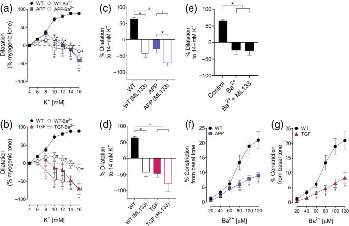
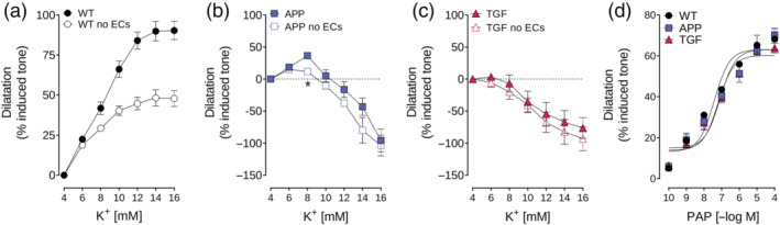
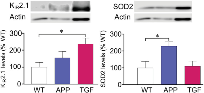
Our findings show that (i) K channel-mediated maximal vasodilatation in APP and TGF mice reaches only 37% and 10%, respectively, of the response seen in WT mice; (ii) K channel dysfunction results from K 2.1 subunit impairment; (iii) about 50% of K -induced artery dilatation is mediated by EC-K channels; (iv) oxidative stress and inflammation impair K channel function, which can be restored by antioxidant and anti-inflammatory drugs; and (v) inflammation induces K 2.1 overexpression and impairs NVC in TGF mice.
Therapies targeting both oxidative stress and inflammation are necessary for full recovery of K 2.1 channel function in cerebrovascular pathology caused by AD and CVD.
内向整流钾(K )通道是神经血管耦合(NVC)中血管舒张的关键效应器。已证实脑内皮细胞(EC)中表达的 K 通道是 NVC 的重要调节剂。阿尔茨海默病(AD)和脑血管疾病(CVD)会影响 EC-K 通道功能,但氧化应激或炎症是否解释了这种损伤仍不清楚。
我们评估了野生型(WT)和过表达人淀粉样前体蛋白突变形式(APP 小鼠,模拟 AD 中观察到的淀粉样 β 诱导的氧化应激)或 TGF-β1 组成型激活形式(TGF 小鼠,模拟脑血管病中观察到的炎症)的转基因小鼠完整和去内皮脑皮层动脉中的 K 通道功能。还研究了抗氧化剂(过氧化氢酶)或抗炎药(吲哚美辛)的益处。体内评估了 NVC 的血管和神经元成分。
我们的发现表明:(i)APP 和 TGF 小鼠中的 K 通道介导的最大血管舒张反应仅分别达到 WT 小鼠反应的 37%和 10%;(ii)K 通道功能障碍是由 K 2.1 亚基损伤引起的;(iii)大约 50%的 K 诱导的动脉舒张是由 EC-K 通道介导的;(iv)氧化应激和炎症会损害 K 通道功能,抗氧化和抗炎药物可以恢复其功能;(v)炎症会导致 TGF 小鼠中 K 2.1 过表达并损害 NVC。
针对氧化应激和炎症的治疗对于 AD 和 CVD 引起的脑血管病中 K 2.1 通道功能的完全恢复是必要的。