Department of Orthopaedic Surgery, Soonchunhyang University Hospital Cheonan, 31, Suncheonhyang 6-gil, Dongam-gu, Cheonan 31151, Korea.
Department of Pathology, Soonchunhyang University Hospital Cheonan, 31, Suncheonhyang 6-gil, Dongam-gu, Cheonan 31151, Korea.
Int J Environ Res Public Health. 2021 Nov 19;18(22):12157. doi: 10.3390/ijerph182212157.
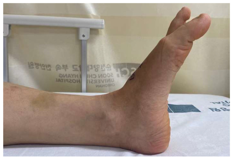
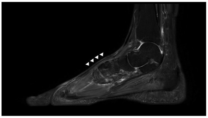
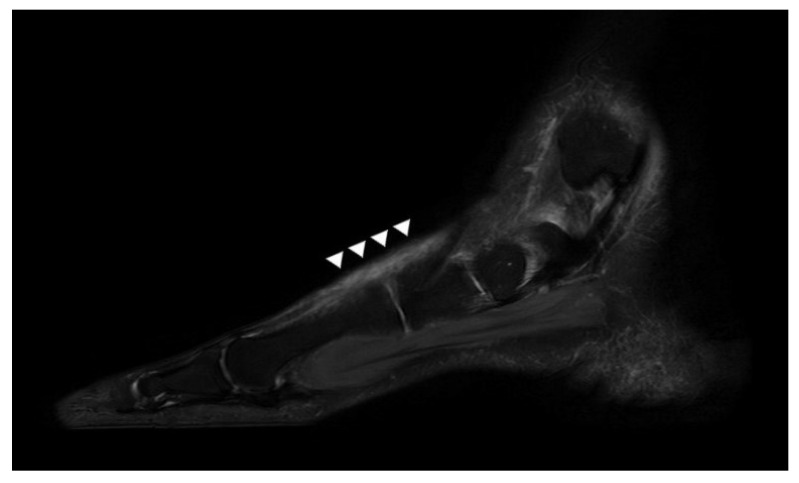
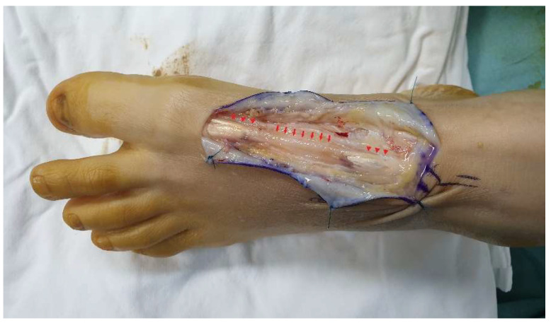
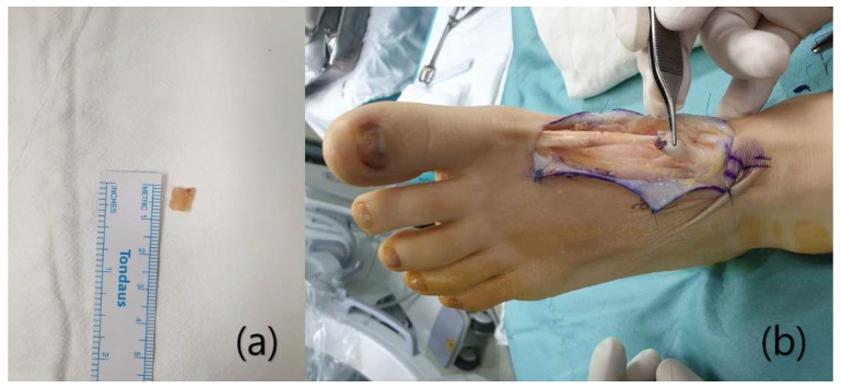
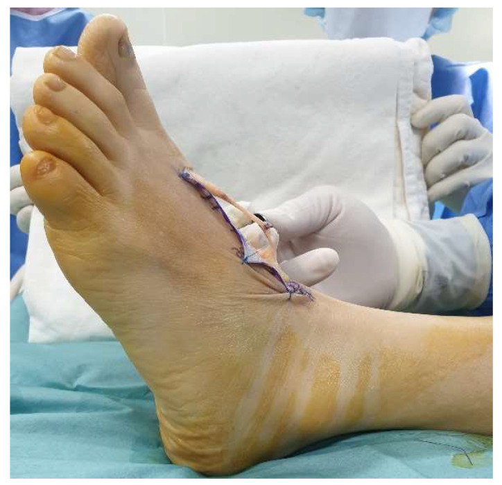
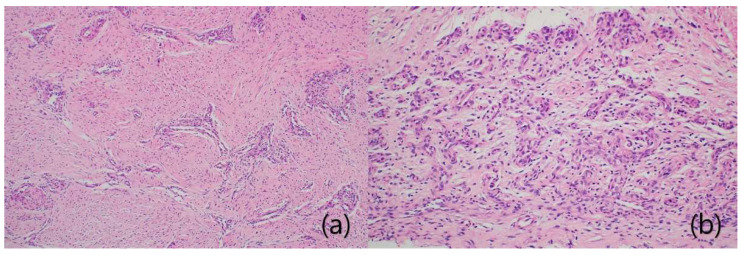
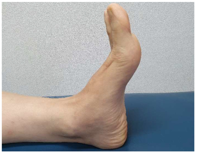
Injury of the extensor hallucis longus (EHL) tendon is relatively rare, but surgical repair is necessary to prevent deformity and gait disturbance. Primary suturing is possible if the condition is acute, but not when it is chronic. The scar tissue between the ruptured ends is a proliferative tissue composed of fibroblasts and collagen fibers. Given the histological similarity to normal tendons, several studies have reported tendon reconstruction using scar tissue. Here, we report a reconstruction of a neglected EHL rupture using interposed scar tissue. A 54-year-old female visited our clinic with a weak extension of a big toe. She had dropped a knife on her foot a month prior, but did not go to hospital. The wound had healed, but she noted dysfunctional extension of the toe and increasing pain. Magnetic resonance imaging (MRI) revealed that EHL continuity was lost and that the proximal tendon stump was displaced toward the midfoot. Scar tissue running in the direction of the original ligament was observed between the ruptured ends. In the surgical field, the scar tissue formed a shape similar to the extensor tendon. Therefore, we performed tendon reconstruction using the interposed scar tissue. For the first 2 postoperative weeks, the ankle and foot were immobilized to protect the repair. Six weeks after surgery, the patient commenced full weight-bearing. At the 3-month follow-up, active extension of the hallux was possible, with a full range of motion. The patient did not feel any discomfort during daily life. Postoperative MRI performed at 1 year revealed that the reconstructed EHL exhibited homogeneously low signal intensity, and was continuous. The AOFAS Hallux Metatarsophalangeal-Interphalangeal scale improved from 57 to 90 points and the FAAM scores improved from 74% to 95% (the Activities of Daily Living subscale) and from 64% to 94% (the Sports subscale). Scar tissue reconstruction is as effective as tendon autografting or allografting, eliminates the risk of donor site morbidity and infection, and requires only a small incision and a short operative time.
踇长伸肌腱(EHL)损伤较为少见,但为防止畸形和步态紊乱,有必要进行手术修复。如果情况是急性的,可以进行直接缝合,但如果是慢性的则不行。断裂两端之间的疤痕组织是由成纤维细胞和胶原纤维组成的增生组织。鉴于其与正常肌腱的组织学相似,已有多项研究报告使用疤痕组织进行肌腱重建。在这里,我们报告了一例使用中间疤痕组织重建被忽视的 EHL 断裂的病例。一位 54 岁的女性因大脚趾无力伸展到我们的诊所就诊。一个月前她在脚上掉了一把刀,但没有去医院。伤口已经愈合,但她注意到脚趾伸展功能障碍且疼痛加剧。磁共振成像(MRI)显示 EHL 连续性丧失,近端肌腱残端向中足移位。在断裂两端之间观察到沿原始韧带方向延伸的疤痕组织。在手术野中,疤痕组织形成了类似于伸肌腱的形状。因此,我们使用中间的疤痕组织进行了肌腱重建。术后 2 周内,踝关节和足部保持固定以保护修复。术后 6 周,患者开始完全负重。术后 3 个月随访时,大脚趾可主动伸展,活动度完全恢复。患者在日常生活中没有感到任何不适。术后 1 年进行的 MRI 显示,重建的 EHL 表现出均匀的低信号强度,且连续性良好。AOFAS 大脚趾跖趾-趾间关节评分从 57 分提高到 90 分,FAAM 评分从 74%提高到 95%(日常生活活动子量表)和 64%提高到 94%(运动子量表)。疤痕组织重建与肌腱自体移植或同种异体移植同样有效,可消除供区发病率和感染的风险,且仅需一个小切口和较短的手术时间。