Zhang Wanxin, Gao Xuemei
Department of Orthodontics, Peking University School and Hospital of Stomatology, Beijing, People's Republic of China.
Int J Womens Health. 2021 Nov 23;13:1129-1137. doi: 10.2147/IJWH.S335728. eCollection 2021.
Menopause is accompanied by a decline in estrogen and progesterone. Several studies have demonstrated that upper airway patency decreases in women after menopause, while morphology changes are still a lack of evidence. This study aimed to explore upper airway morphology changes in perimenopausal and postmenopausal women.
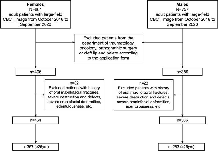
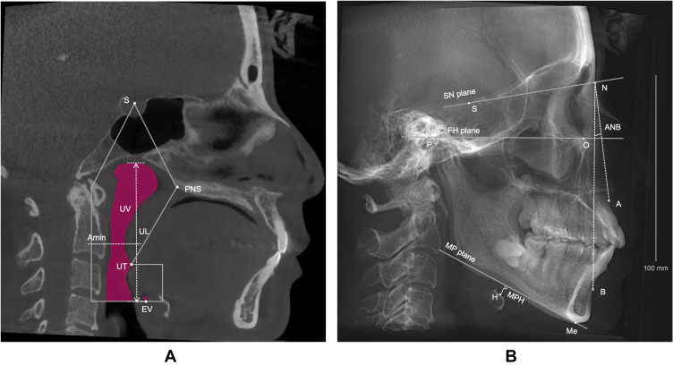
This retrospective cross-sectional study included 367 consecutive Chinese female patients over 25 years old who had routinely taken large-field cone beam computed tomography in the imaging library of Peking University School and Hospital of Stomatology from October 2016 to September 2020. A total of 283 males were screened as sex controls according to the same age group. Upper airway morphology, hyoid position and facial pattern were measured. The association between perimenopausal and postmenopausal years and upper airway morphology in both sexes was analyzed.
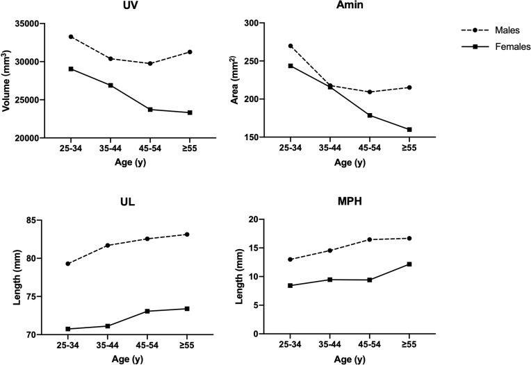
Perimenopausal women (aged 45-54 years) showed a significant decrease in the volume (3172.91mm, 95% CI = 653.86-5691.96) and minimum cross-sectional area (37.08 mm, 95% CI = 5.36-68.80), and a significant increase in the length (-1.96mm, 95% CI = -3.62 to -0.29) of upper airway compared to adjacent reproductive years (aged 35-44), while this difference was neither seen in other adjacent two reproductive age groups of females nor in the same age groups of males. In postmenopausal women (55 years and older), hyoid position was significantly lower (-2.74mm, 95% CI = -4.42 to -1.07) than either age group, while no similar changes were seen in men.
Women had smaller airway volume, reduced upper airway cross-sectional area and longer airway length in perimenopausal years, and a significantly lower hyoid position in postmenopausal years. These changes may be related to menopause itself and independent of the changes associated with aging.
绝经伴随着雌激素和孕激素水平的下降。多项研究表明,绝经后女性上气道通畅性降低,而形态学变化仍缺乏证据。本研究旨在探讨围绝经期和绝经后女性的上气道形态变化。
这项回顾性横断面研究纳入了2016年10月至2020年9月期间在北京大学口腔医院影像库中常规接受大视野锥形束计算机断层扫描的367例年龄超过25岁的中国女性患者。根据相同年龄组筛选出283名男性作为性别对照。测量上气道形态、舌骨位置和面部形态。分析围绝经期和绝经后年限与两性上气道形态之间的关联。
围绝经期女性(45 - 54岁)与相邻生育期(35 - 44岁)相比,上气道容积显著减小(3172.91mm³,95%可信区间 = 653.86 - 5691.96),最小横截面积显著减小(37.08mm²,95%可信区间 = 5.36 - 68.80),气道长度显著增加(-1.96mm,95%可信区间 = -3.62至 -0.29),而在其他相邻两个生育年龄组的女性以及同年龄组的男性中均未观察到这种差异。绝经后女性(55岁及以上)舌骨位置显著低于其他任何年龄组,而男性未出现类似变化。
女性在围绝经期气道容积较小、上气道横截面积减小且气道长度增加,在绝经后舌骨位置显著降低。这些变化可能与绝经本身有关,且独立于与衰老相关的变化。