Research Institute of Atherosclerotic Disease, Xi'an Jiaotong University Cardiovascular Research Centre, Xi'an, Shaanxi 710061, China.
Laboratory Animal Center, Xi'an Jiaotong University Health Science Centre, Xi'an, Shaanxi 710061, China.
Oxid Med Cell Longev. 2021 Nov 22;2021:3010577. doi: 10.1155/2021/3010577. eCollection 2021.
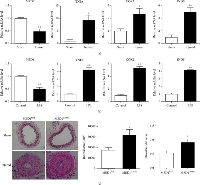
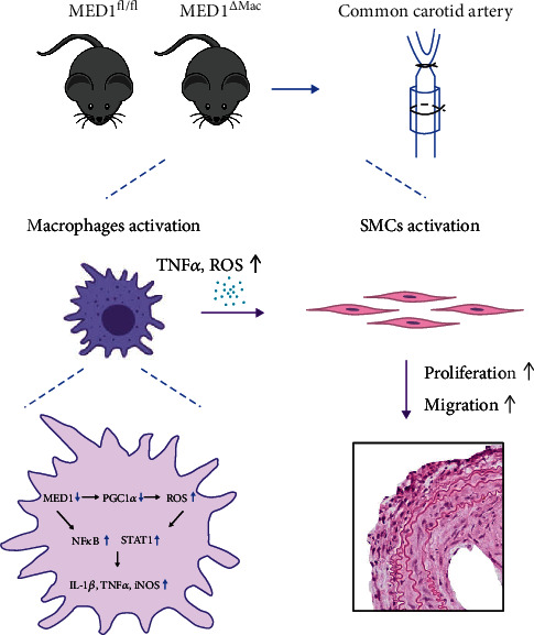
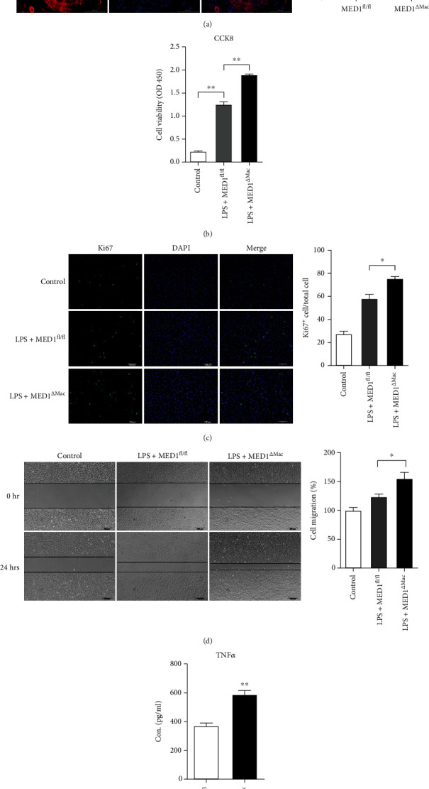
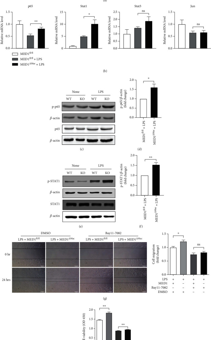
Mediator complex subunit 1 (MED1) is a component of the mediator complex and functions as a coactivator involved in the regulated transcription of nearly all RNA polymerase II-dependent genes. Previously, we showed that MED1 in macrophages has a protective effect on atherosclerosis; however, the effect of MED1 on intimal hyperplasia and mechanisms regulating proinflammatory cytokine production after macrophage MED1 deletion are still unknown. In this study, we report that MED1 macrophage-specific knockout (MED1 ) mice showed aggravated neointimal hyperplasia, vascular smooth muscle cells (VSMCs), and macrophage accumulation in injured arteries. Moreover, MED1 mice showed increased proinflammatory cytokine production after an injury to the artery. After lipopolysaccharide (LPS) treatment, MED1 macrophages showed increased generation of reactive oxygen species (ROS) and reduced expression of peroxisome proliferative activated receptor gamma coactivator-1 (PGC1) and antioxidant enzymes, including catalase and glutathione reductase. The overexpression of PGC1 attenuated the effects of MED1 deficiency in macrophages. , conditioned media from MED1 macrophages induced more proliferation and migration of VSMCs. To explore the potential mechanisms by which MED1 affects inflammation, macrophages were treated with BAY11-7082 before LPS treatment, and the results showed that MED1 macrophages exhibited increased expression of phosphorylated-p65 and phosphorylated signal transducer and activator of transcription 1 (p-STAT1) compared with the control macrophages, suggesting the enhanced activation of NF-B and STAT1. In summary, these data showed that MED1 deficiency enhanced inflammation and the proliferation and migration of VSMCs in injured vascular tissue, which may result from the activation of NF-B and STAT1 due to the accumulation of ROS.
中介复合物亚基 1(MED1)是中介复合物的一个组成部分,作为一种共激活因子发挥作用,参与几乎所有 RNA 聚合酶 II 依赖性基因的调节转录。先前,我们表明巨噬细胞中的 MED1 对动脉粥样硬化具有保护作用;然而,MED1 对内膜增生的影响以及调节巨噬细胞 MED1 缺失后促炎细胞因子产生的机制仍不清楚。在这项研究中,我们报告了 MED1 巨噬细胞特异性敲除(MED1)小鼠在损伤动脉中表现出加重的新生内膜增生、血管平滑肌细胞(VSMCs)和巨噬细胞积聚。此外,MED1 小鼠在动脉损伤后表现出促炎细胞因子产生增加。在脂多糖(LPS)处理后,MED1 巨噬细胞表现出活性氧(ROS)生成增加和过氧化物酶体增殖物激活受体γ共激活因子 1(PGC1)和抗氧化酶,包括过氧化氢酶和谷胱甘肽还原酶表达减少。PGC1 的过表达减弱了 MED1 缺乏对巨噬细胞的影响。 ,来自 MED1 巨噬细胞的条件培养基诱导 VSMCs 更多的增殖和迁移。为了探索 MED1 影响炎症的潜在机制,用 BAY11-7082 处理巨噬细胞后用 LPS 处理,结果表明与对照巨噬细胞相比,MED1 巨噬细胞表现出磷酸化-p65 和磷酸化信号转导和转录激活因子 1(p-STAT1)的表达增加,表明 NF-B 和 STAT1 的激活增强。总之,这些数据表明,MED1 缺乏增强了损伤血管组织中的炎症以及 VSMCs 的增殖和迁移,这可能是由于 ROS 积累导致 NF-B 和 STAT1 的激活所致。