Yamada Shuhei, Kijima Noriyuki, Nakagawa Tomoyoshi, Hirayama Ryuichi, Kinoshita Manabu, Kagawa Naoki, Kishima Haruhiko
Department of Neurosurgery, Osaka University Graduate School of Medicine, Suita, Japan.
Front Neurol. 2021 Nov 17;12:769656. doi: 10.3389/fneur.2021.769656. eCollection 2021.
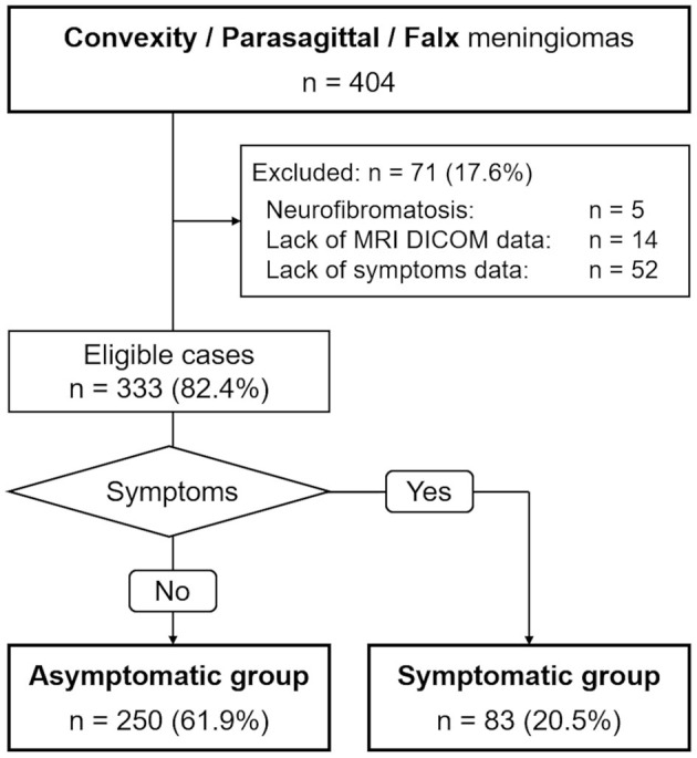
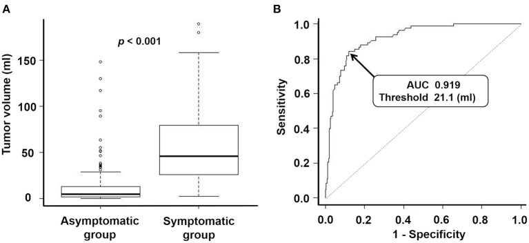
Meningiomas are the most common primary intracranial neoplasms and clinical symptom appearance depends on their volume and location. This study aimed to identify factors that influence clinical symptoms and to determine a specific threshold tumor volume for the prediction of symptomatic progression in patients with convexity, parasagittal, and falx meningiomas. We retrospectively studied patients with radiologically suspected convexity, parasagittal, or falx meningiomas at our institution. The data of three hundred thirty-three patients were analyzed. We further divided patients into two groups based on clinical symptoms: an asymptomatic group (250 cases) and a symptomatic group (83 cases). Univariate analysis revealed significant differences between the groups in terms of sex ( = 0.002), age at the time of volumetric analysis ( < 0.001), hyperintense lesions on T2-weighted images ( = 0.029), peritumoral edema ( < 0.001), maximum tumor diameter ( < 0.001), and tumor volume ( < 0.001). Further multivariate analysis revealed significant differences between the groups in terms of age at the time of volumetric analysis ( = 0.002), peritumoral edema ( < 0.001), and tumor volume ( < 0.001). The receiver operating characteristic curve revealed a threshold tumor volume of 21.1 ml for predicting whether a patient would develop symptoms (sensitivity 0.843, specificity 0.880, an area under the curve 0.919 [95% confidence interval: 0.887-0.951]). We identified factors predictive of clinical symptoms in patients with convexity, parasagittal, and falx meningiomas and determined the first-ever threshold tumor volume for predicting symptomatic progression in such patients.
脑膜瘤是最常见的原发性颅内肿瘤,其临床症状的出现取决于肿瘤的体积和位置。本研究旨在确定影响临床症状的因素,并确定预测凸面、矢状窦旁和镰旁脑膜瘤患者症状进展的特定肿瘤体积阈值。我们对本院放射学怀疑为凸面、矢状窦旁或镰旁脑膜瘤的患者进行了回顾性研究。分析了333例患者的数据。我们根据临床症状将患者进一步分为两组:无症状组(250例)和有症状组(83例)。单因素分析显示,两组在性别(P = 0.002)、体积分析时的年龄(P < 0.001)、T2加权图像上的高信号病变(P = 0.029)、瘤周水肿(P < 0.001)、最大肿瘤直径(P < 0.001)和肿瘤体积(P < 0.001)方面存在显著差异。进一步的多因素分析显示,两组在体积分析时的年龄(P = 0.002)、瘤周水肿(P < 0.001)和肿瘤体积(P < 0.001)方面存在显著差异。受试者工作特征曲线显示,预测患者是否会出现症状的肿瘤体积阈值为21.1 ml(敏感性0.843,特异性0.880,曲线下面积0.919 [95%置信区间:0.887 - 0.951])。我们确定了凸面、矢状窦旁和镰旁脑膜瘤患者临床症状的预测因素,并确定了此类患者症状进展预测的首个肿瘤体积阈值。