• 文献检索
  • 文档翻译
  • 深度研究
  • 学术资讯
  • Suppr Zotero 插件Zotero 插件
  • 邀请有礼
  • 套餐&价格
  • 历史记录
应用&插件
Suppr Zotero 插件Zotero 插件浏览器插件Mac 客户端Windows 客户端微信小程序
定价
高级版会员购买积分包购买API积分包
服务
文献检索文档翻译深度研究API 文档MCP 服务
关于我们
关于 Suppr公司介绍联系我们用户协议隐私条款
关注我们

Suppr 超能文献

核心技术专利:CN118964589B侵权必究
粤ICP备2023148730 号-1Suppr @ 2026

文献检索

告别复杂PubMed语法,用中文像聊天一样搜索,搜遍4000万医学文献。AI智能推荐,让科研检索更轻松。

立即免费搜索

文件翻译

保留排版,准确专业,支持PDF/Word/PPT等文件格式,支持 12+语言互译。

免费翻译文档

深度研究

AI帮你快速写综述,25分钟生成高质量综述,智能提取关键信息,辅助科研写作。

立即免费体验

细胞外囊泡分子特征可表征卵巢癌转移动态性

Extracellular Vesicle Molecular Signatures Characterize Metastatic Dynamicity in Ovarian Cancer.

作者信息

Gonda Amber, Zhao Nanxia, Shah Jay V, Siebert Jake N, Gunda Srujanesh, Inan Berk, Kwon Mijung, Libutti Steven K, Moghe Prabhas V, Francis Nicola L, Ganapathy Vidya

机构信息

Department of Biomedical Engineering, Rutgers University, Piscataway, NJ, United States.

Department of Chemical and Biochemical Engineering, Rutgers University, Piscataway, NJ, United States.

出版信息

Front Oncol. 2021 Nov 18;11:718408. doi: 10.3389/fonc.2021.718408. eCollection 2021.

DOI:10.3389/fonc.2021.718408
PMID:34868914
原文链接:https://pmc.ncbi.nlm.nih.gov/articles/PMC8637407/
Abstract

BACKGROUND

Late-stage diagnosis of ovarian cancer, a disease that originates in the ovaries and spreads to the peritoneal cavity, lowers 5-year survival rate from 90% to 30%. Early screening tools that can: i) detect with high specificity and sensitivity before conventional tools such as transvaginal ultrasound and CA-125, ii) use non-invasive sampling methods and iii) longitudinally significantly increase survival rates in ovarian cancer are needed. Studies that employ blood-based screening tools using circulating tumor-cells, -DNA, and most recently tumor-derived small extracellular vesicles (sEVs) have shown promise in non-invasive detection of cancer before standard of care. Our findings in this study show the promise of a sEV-derived signature as a non-invasive longitudinal screening tool in ovarian cancer.

METHODS

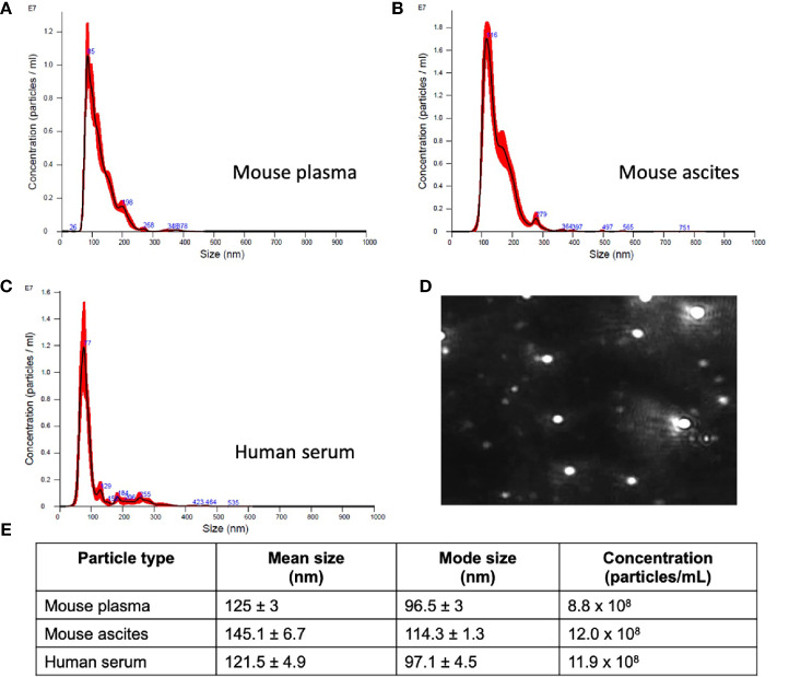
Human serum samples as well as plasma and ascites from a mouse model of ovarian cancer were collected at various disease stages. Small extracellular vesicles (sEVs) were extracted using a commercially available kit. RNA was isolated from lysed sEVs, and quantitative RT-PCR was performed to identify specific metastatic gene expression.

CONCLUSION

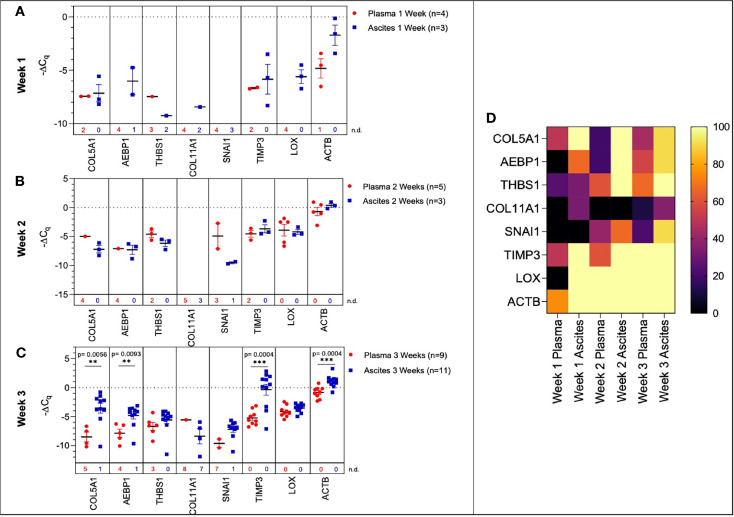
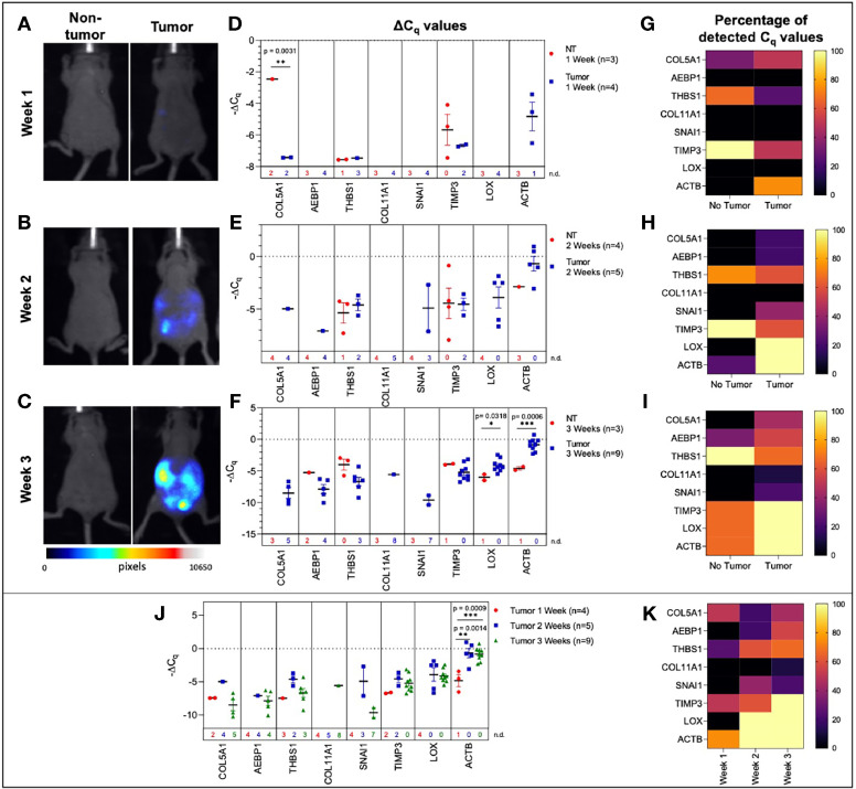
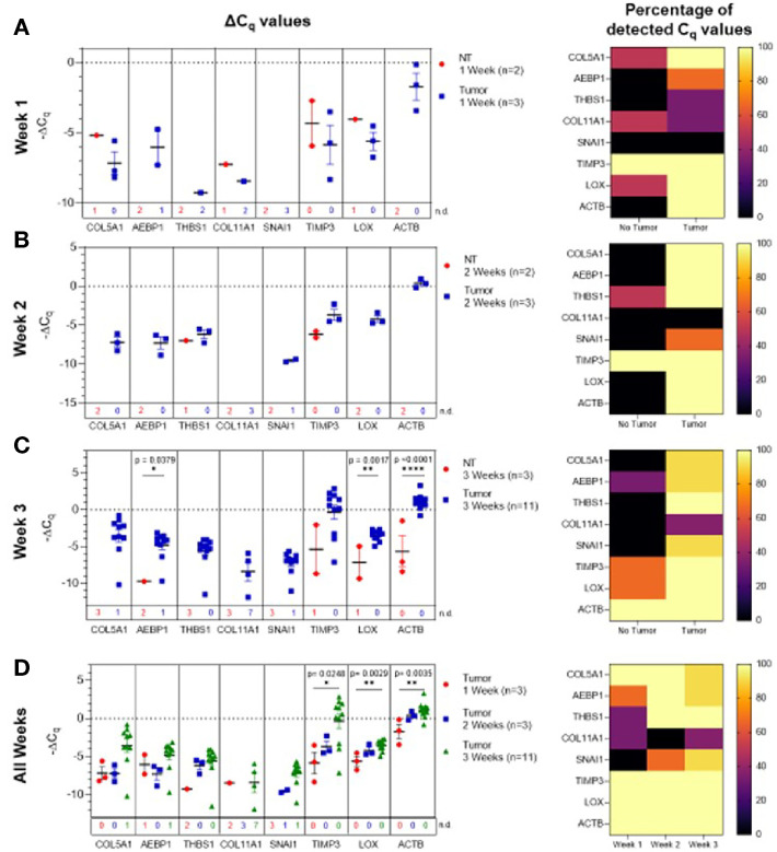
This paper highlights the potential of sEVs in monitoring ovarian cancer progression and metastatic development. We identified a 7-gene panel in sEVs derived from plasma, serum, and ascites that overlapped with an established metastatic ovarian carcinoma signature. We found the 7-gene panel to be differentially expressed with tumor development and metastatic spread in a mouse model of ovarian cancer. The most notable finding was a significant change in the ascites-derived sEV gene signature that overlapped with that of the plasma-derived sEV signature at varying stages of disease progression. While there were quantifiable changes in genes from the 7-gene panel in serum-derived sEVs from ovarian cancer patients, we were unable to establish a definitive signature due to low sample number. Taken together our findings show that differential expression of metastatic genes derived from circulating sEVs present a minimally invasive screening tool for ovarian cancer detection and longitudinal monitoring of molecular changes associated with progression and metastatic spread.

摘要

背景

卵巢癌起源于卵巢并扩散至腹腔,晚期诊断会使5年生存率从90%降至30%。因此,需要一种早期筛查工具,该工具能够:i)在经阴道超声和CA - 125等传统工具之前进行高特异性和高灵敏度检测;ii)采用非侵入性采样方法;iii)在卵巢癌中显著提高纵向生存率。使用基于血液的筛查工具,检测循环肿瘤细胞、肿瘤DNA以及最近的肿瘤来源小细胞外囊泡(sEV)的研究,在癌症的非侵入性检测方面已显示出优于标准护理的前景。我们在本研究中的发现表明,sEV衍生的标志物有望作为卵巢癌的非侵入性纵向筛查工具。

方法

在不同疾病阶段收集人血清样本以及卵巢癌小鼠模型的血浆和腹水。使用市售试剂盒提取小细胞外囊泡(sEV)。从裂解的sEV中分离RNA,并进行定量RT - PCR以鉴定特定转移基因的表达。

结论

本文强调了sEV在监测卵巢癌进展和转移发展方面的潜力。我们在源自血浆、血清和腹水的sEV中鉴定出一个7基因组合,该组合与已确立的转移性卵巢癌标志物重叠。我们发现该7基因组合在卵巢癌小鼠模型中随肿瘤发展和转移扩散而差异表达。最显著的发现是,在疾病进展的不同阶段,腹水中衍生的sEV基因标志物与血浆中衍生的sEV标志物发生了显著变化。虽然卵巢癌患者血清中衍生的sEV的7基因组合中的基因存在可量化变化,但由于样本数量少,我们无法确定一个明确的标志物。综上所述,我们的研究结果表明,循环sEV衍生的转移基因的差异表达为卵巢癌检测以及与进展和转移扩散相关的分子变化的纵向监测提供了一种微创筛查工具。

https://cdn.ncbi.nlm.nih.gov/pmc/blobs/22ee/8637407/99061643426c/fonc-11-718408-g006.jpg
https://cdn.ncbi.nlm.nih.gov/pmc/blobs/22ee/8637407/b92e4d1fac09/fonc-11-718408-g001.jpg
https://cdn.ncbi.nlm.nih.gov/pmc/blobs/22ee/8637407/2f8f9ed6f192/fonc-11-718408-g002.jpg
https://cdn.ncbi.nlm.nih.gov/pmc/blobs/22ee/8637407/ee1b66f1a9a8/fonc-11-718408-g003.jpg
https://cdn.ncbi.nlm.nih.gov/pmc/blobs/22ee/8637407/735a7ecef21d/fonc-11-718408-g004.jpg
https://cdn.ncbi.nlm.nih.gov/pmc/blobs/22ee/8637407/9668cd59cb93/fonc-11-718408-g005.jpg
https://cdn.ncbi.nlm.nih.gov/pmc/blobs/22ee/8637407/99061643426c/fonc-11-718408-g006.jpg

相似文献

1
Extracellular Vesicle Molecular Signatures Characterize Metastatic Dynamicity in Ovarian Cancer.细胞外囊泡分子特征可表征卵巢癌转移动态性
Front Oncol. 2021 Nov 18;11:718408. doi: 10.3389/fonc.2021.718408. eCollection 2021.
2
Lactadherin immunoblockade in small extracellular vesicles inhibits sEV-mediated increase of pro-metastatic capacities.乳铁蛋白免疫阻断小细胞外囊泡抑制 sEV 介导的促转移能力增加。
Biol Res. 2024 Jan 3;57(1):1. doi: 10.1186/s40659-023-00477-8.
3
Protein Panel of Serum-Derived Small Extracellular Vesicles for the Screening and Diagnosis of Epithelial Ovarian Cancer.用于上皮性卵巢癌筛查和诊断的血清来源小细胞外囊泡蛋白质组
Cancers (Basel). 2022 Jul 30;14(15):3719. doi: 10.3390/cancers14153719.
4
microRNA-141-3p-containing small extracellular vesicles derived from epithelial ovarian cancer cells promote endothelial cell angiogenesis through activating the JAK/STAT3 and NF-κB signaling pathways.源自上皮性卵巢癌细胞的含微小RNA-141-3p的小细胞外囊泡通过激活JAK/STAT3和NF-κB信号通路促进内皮细胞血管生成。
J Cell Commun Signal. 2020 Jun;14(2):233-244. doi: 10.1007/s12079-020-00548-5. Epub 2020 Feb 7.
5
Liver injury changes the biological characters of serum small extracellular vesicles and reprograms hepatic macrophages in mice.肝损伤改变了血清中小细胞外囊泡的生物学特性,并重塑了小鼠肝巨噬细胞。
World J Gastroenterol. 2021 Nov 21;27(43):7509-7529. doi: 10.3748/wjg.v27.i43.7509.
6
Predicting chemoresponsiveness in epithelial ovarian cancer patients using circulating small extracellular vesicle-derived plasma gelsolin.使用循环小细胞外囊泡衍生的血浆gelsolin 预测上皮性卵巢癌患者的化疗反应性。
J Ovarian Res. 2023 Jan 16;16(1):14. doi: 10.1186/s13048-022-01086-x.
7
Pharmacokinetic Approach for the Elucidation of Elevated Plasma Small Extracellular Vesicle (sEV) Concentration in Cancer.阐明癌症患者血浆中小细胞外囊泡(sEV)浓度升高的药代动力学方法。
J Pharm Sci. 2023 Jul;112(7):1967-1974. doi: 10.1016/j.xphs.2023.03.017. Epub 2023 Mar 30.
8
Improving SERS biosensors for the analysis of ovarian cancer-derived small extracellular vesicles.用于分析卵巢癌来源的小细胞外囊泡的 SERS 生物传感器的改进。
Analyst. 2023 Jun 26;148(13):3074-3086. doi: 10.1039/d3an00398a.
9
ISG15 mediates the function of extracellular vesicles in promoting ovarian cancer progression and metastasis.ISG15介导细胞外囊泡在促进卵巢癌进展和转移中的功能。
J Extracell Biol. 2024 Jan 31;3(2):e92. doi: 10.1002/jex2.92. eCollection 2024 Feb.
10
Plasmonic nano-aperture label-free imaging of single small extracellular vesicles for cancer detection.用于癌症检测的单个小细胞外囊泡的表面等离子体纳米孔无标记成像
Commun Med (Lond). 2024 May 25;4(1):100. doi: 10.1038/s43856-024-00514-x.

引用本文的文献

1
An Update of Extracellular Vesicle Involvement in Different Steps of Cancer Metastasis and Targeting Strategies.细胞外囊泡在癌症转移不同步骤中的作用及靶向策略的研究进展。
Curr Med Chem. 2024;31(28):4495-4509. doi: 10.2174/0109298673273299231121044055.
2
Simultaneously targeting extracellular vesicle trafficking and TGF-β receptor kinase activity blocks signaling hyperactivation and metastasis.同时靶向细胞外囊泡转运和 TGF-β 受体激酶活性可阻断信号过度激活和转移。
Signal Transduct Target Ther. 2023 Dec 18;8(1):456. doi: 10.1038/s41392-023-01711-1.
3
Extracellular vesicle small RNA cargo discriminates non-cancer donors from pediatric B-lymphoblastic leukemia patients.

本文引用的文献

1
Overexpressed COL5A1 is correlated with tumor progression, paclitaxel resistance, and tumor-infiltrating immune cells in ovarian cancer.COL5A1 过表达与卵巢癌的肿瘤进展、紫杉醇耐药和肿瘤浸润免疫细胞相关。
J Cell Physiol. 2021 Oct;236(10):6907-6919. doi: 10.1002/jcp.30350. Epub 2021 Mar 3.
2
Exosome-based liquid biopsies in cancer: opportunities and challenges.基于外泌体的癌症液体活检:机遇与挑战。
Ann Oncol. 2021 Apr;32(4):466-477. doi: 10.1016/j.annonc.2021.01.074. Epub 2021 Feb 4.
3
CA125 and Ovarian Cancer: A Comprehensive Review.
细胞外囊泡小RNA货物可区分非癌症捐赠者与小儿B淋巴细胞白血病患者。
Front Oncol. 2023 Nov 13;13:1272883. doi: 10.3389/fonc.2023.1272883. eCollection 2023.
4
Extracellular Vesicles-ceRNAs as Ovarian Cancer Biomarkers: Looking into circRNA-miRNA-mRNA Code.作为卵巢癌生物标志物的细胞外囊泡-ceRNAs:探究环状RNA-微小RNA-信使RNA编码
Cancers (Basel). 2022 Jul 13;14(14):3404. doi: 10.3390/cancers14143404.
CA125与卵巢癌:一篇综述
Cancers (Basel). 2020 Dec 11;12(12):3730. doi: 10.3390/cancers12123730.
4
Stromal POSTN induced by TGF-β1 facilitates the migration and invasion of ovarian cancer.TGF-β1 诱导的基质 POSTN 促进卵巢癌细胞的迁移和侵袭。
Gynecol Oncol. 2021 Feb;160(2):530-538. doi: 10.1016/j.ygyno.2020.11.026. Epub 2020 Dec 13.
5
Concepts of extracellular matrix remodelling in tumour progression and metastasis.肿瘤进展和转移中外细胞基质重塑的概念。
Nat Commun. 2020 Oct 9;11(1):5120. doi: 10.1038/s41467-020-18794-x.
6
Collagen-rich omentum is a premetastatic niche for integrin α2-mediated peritoneal metastasis.富含胶原蛋白的网膜是整合素 α2 介导的腹膜转移的前转移生态位。
Elife. 2020 Oct 7;9:e59442. doi: 10.7554/eLife.59442.
7
Prognostic significance of abnormal matrix collagen remodeling in colorectal cancer based on histologic and bioinformatics analysis.基于组织学和生物信息学分析的结直肠癌异常基质胶原重塑的预后意义。
Oncol Rep. 2020 Oct;44(4):1671-1685. doi: 10.3892/or.2020.7729. Epub 2020 Aug 11.
8
Molecular mediators of peritoneal metastasis in pancreatic cancer.胰腺癌腹膜转移的分子介质。
Cancer Metastasis Rev. 2020 Dec;39(4):1223-1243. doi: 10.1007/s10555-020-09924-4. Epub 2020 Aug 11.
9
Non-invasive early detection of cancer four years before conventional diagnosis using a blood test.利用血液检测,可在常规诊断前四年进行非侵入性的癌症早期检测。
Nat Commun. 2020 Jul 21;11(1):3475. doi: 10.1038/s41467-020-17316-z.
10
AEBP1 is a Novel Oncogene: Mechanisms of Action and Signaling Pathways.AEBP1是一种新型癌基因:作用机制与信号通路
J Oncol. 2020 May 27;2020:8097872. doi: 10.1155/2020/8097872. eCollection 2020.