Uchikado Yoshihiro, Ikeda Yoshiyuki, Sasaki Yuichi, Iwabayashi Masaaki, Akasaki Yuichi, Ohishi Mitsuru
Department of Cardiovascular Medicine and Hypertension, Graduate School of Medical and Dental Sciences Kagoshima University, Kagoshima, Japan.
Front Cardiovasc Med. 2021 Nov 17;8:788655. doi: 10.3389/fcvm.2021.788655. eCollection 2021.
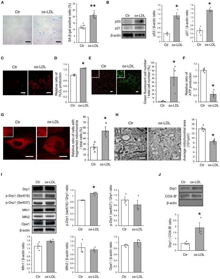
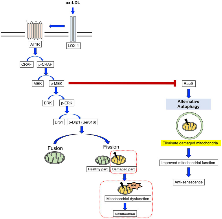
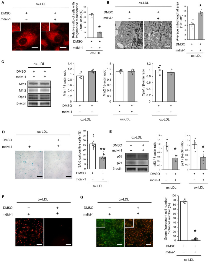
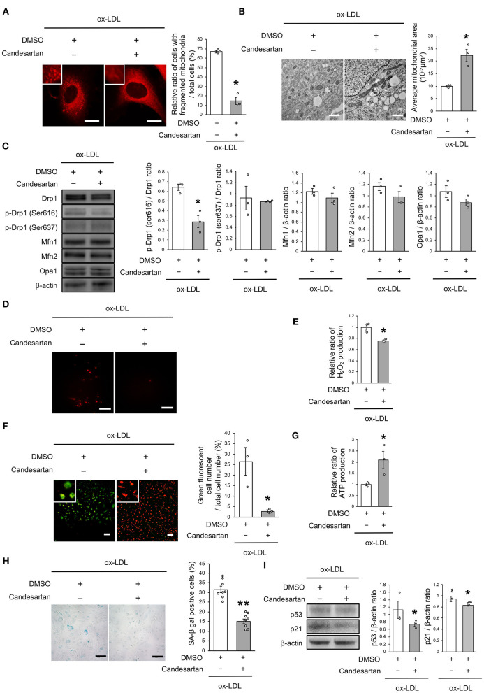
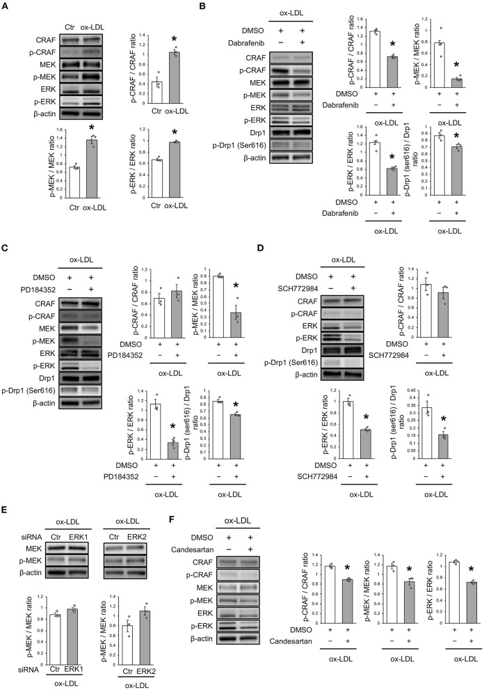
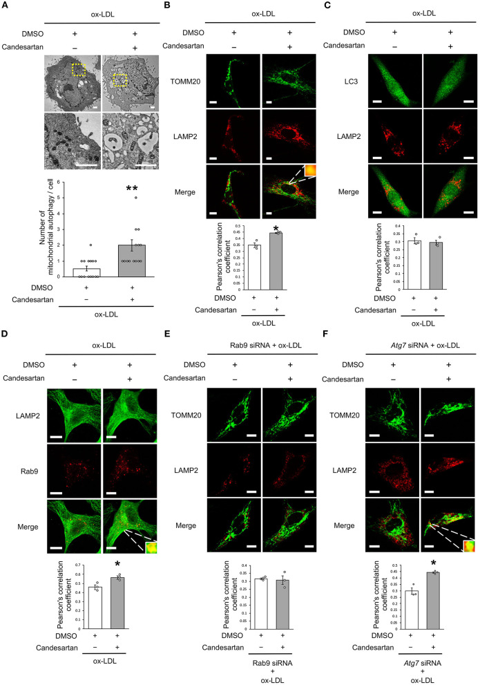
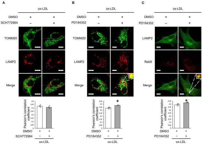
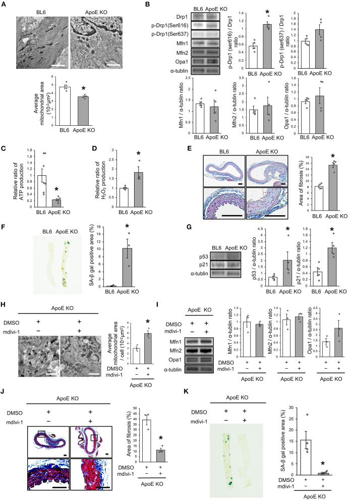
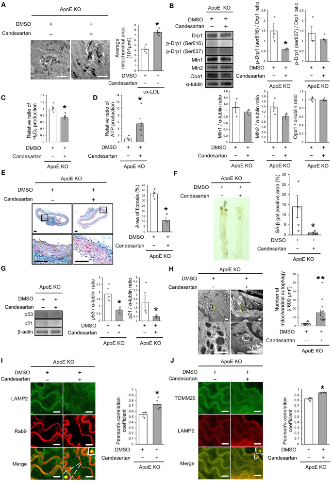
Lectin-like oxidized low-density lipoprotein (ox-LDL) causes vascular senescence and atherosclerosis. It has been reported that ox-LDL scavenger receptor-1 (LOX-1) is associated with the angiotensin II type 1 receptor (AT1R). While mitochondria play a crucial role in the development of vascular senescence and atherosclerosis, they also undergo quality control through mitochondrial dynamics and autophagy. The aim of this study was to investigate (1) whether LOX-1 associates with AT1R, (2) if this regulates mitochondrial quality control, and (3) whether AT1R inhibition using Candesartan might ameliorate ox-LDL-induced vascular senescence. We performed and experiments using vascular smooth muscle cells (VSMCs), and C57BL/6 and apolipoprotein E-deficient (ApoE KO) mice. Administration of oxidized low-density lipoprotein (ox-LDL) to VSMCs induced mitochondrial dysfunction and cellular senescence accompanied by excessive mitochondrial fission, due to the activation of fission factor Drp1, which was derived from the activation of the Raf/MEK/ERK pathway. Administration of either Drp1 inhibitor, mdivi-1, or AT1R blocker candesartan attenuated these alterations. Electron microscopy and immunohistochemistry of the co-localization of LAMP2 with TOMM20 signal showed that AT1R inhibition also increased mitochondrial autophagy, but this was not affected by Atg7 deficiency. Conversely, AT1R inhibition increased the co-localization of LAMP2 with Rab9 signal. Moreover, AT1R inhibition-induced mitochondrial autophagy was abolished by Rab9 deficiency, suggesting that AT1R signaling modulated mitochondrial autophagy derived from Rab9-dependent alternative autophagy. Inhibition of the Raf/MEK/ERK pathway also decreased the excessive mitochondrial fission, and Rab9-dependent mitochondrial autophagy, suggesting that AT1R signaling followed the Raf/MEK/ERK axis modulated both mitochondrial dynamics and autophagy. The degree of mitochondrial dysfunction, reactive oxygen species production, vascular senescence, atherosclerosis, and the number of fragmented mitochondria accompanied by Drp1 activation were all higher in ApoE KO mice than in C57BL/6 mice. These detrimental alterations were successfully restored, and mitochondrial autophagy was upregulated with the administration of candesartan to ApoE KO mice. The association of LOX-1 with AT1R was found to play a crucial role in regulating mitochondrial quality control, as cellular/vascular senescence is induced by ox-LDL, and AT1R inhibition improves the adverse effects of ox-LDL.
凝集素样氧化低密度脂蛋白(ox-LDL)可导致血管衰老和动脉粥样硬化。据报道,ox-LDL清道夫受体-1(LOX-1)与血管紧张素II 1型受体(AT1R)有关。虽然线粒体在血管衰老和动脉粥样硬化的发展中起着关键作用,但它们也通过线粒体动力学和自噬进行质量控制。本研究的目的是调查:(1)LOX-1是否与AT1R相关;(2)这是否调节线粒体质量控制;(3)使用坎地沙坦抑制AT1R是否可以改善ox-LDL诱导的血管衰老。我们使用血管平滑肌细胞(VSMC)以及C57BL/6和载脂蛋白E缺陷(ApoE KO)小鼠进行了实验。向VSMC中给予氧化低密度脂蛋白(ox-LDL)会诱导线粒体功能障碍和细胞衰老,并伴有过度的线粒体分裂,这是由于裂变因子Drp1的激活所致,而Drp1的激活源于Raf/MEK/ERK途径的激活。给予Drp1抑制剂mdivi-1或AT1R阻滞剂坎地沙坦可减轻这些改变。电子显微镜检查以及LAMP2与TOMM20信号共定位的免疫组织化学显示,抑制AT1R也会增加线粒体自噬,但这不受Atg7缺陷的影响。相反,抑制AT1R会增加LAMP2与Rab9信号的共定位。此外,Rab9缺陷可消除AT1R抑制诱导的线粒体自噬,这表明AT1R信号传导调节了源自Rab9依赖性替代自噬的线粒体自噬。抑制Raf/MEK/ERK途径也会减少过度的线粒体分裂以及Rab9依赖性线粒体自噬,这表明遵循Raf/MEK/ERK轴的AT1R信号传导调节了线粒体动力学和自噬。ApoE KO小鼠中线粒体功能障碍的程度、活性氧的产生、血管衰老、动脉粥样硬化以及伴有Drp1激活的线粒体片段化数量均高于C57BL/6小鼠。通过给ApoE KO小鼠施用坎地沙坦,这些有害改变得以成功恢复,并且线粒体自噬上调。发现LOX-1与AT1R的关联在调节线粒体质量控制中起关键作用,因为ox-LDL会诱导细胞/血管衰老,而抑制AT1R可改善ox-LDL的不良影响。