• 文献检索
  • 文档翻译
  • 深度研究
  • 学术资讯
  • Suppr Zotero 插件Zotero 插件
  • 邀请有礼
  • 套餐&价格
  • 历史记录
应用&插件
Suppr Zotero 插件Zotero 插件浏览器插件Mac 客户端Windows 客户端微信小程序
定价
高级版会员购买积分包购买API积分包
服务
文献检索文档翻译深度研究API 文档MCP 服务
关于我们
关于 Suppr公司介绍联系我们用户协议隐私条款
关注我们

Suppr 超能文献

核心技术专利:CN118964589B侵权必究
粤ICP备2023148730 号-1Suppr @ 2026

文献检索

告别复杂PubMed语法,用中文像聊天一样搜索,搜遍4000万医学文献。AI智能推荐,让科研检索更轻松。

立即免费搜索

文件翻译

保留排版,准确专业,支持PDF/Word/PPT等文件格式,支持 12+语言互译。

免费翻译文档

深度研究

AI帮你快速写综述,25分钟生成高质量综述,智能提取关键信息,辅助科研写作。

立即免费体验

[F]氟代阿尔伐斯汀环丙基非肽类 FAPI-04 正电子发射断层扫描/计算机断层扫描(PET/CT)摄取在转移灶的 PET/CT 成像上可能有助于鉴别不同病理类型的肺癌。

[F]AlF-NOTA-FAPI-04 PET/CT uptake in metastatic lesions on PET/CT imaging might distinguish different pathological types of lung cancer.

机构信息

Cheeloo College of Medicine, Shandong University, Jinan, China.

Department of Radiology, Shandong Cancer Hospital and Institute, Shandong First Medical University, Shandong Academy of Medical Sciences, Jinan, 250117, Shandong, China.

出版信息

Eur J Nucl Med Mol Imaging. 2022 Apr;49(5):1671-1681. doi: 10.1007/s00259-021-05638-z. Epub 2021 Dec 6.

DOI:10.1007/s00259-021-05638-z
PMID:34870727
原文链接:https://pmc.ncbi.nlm.nih.gov/articles/PMC8940861/
Abstract

PURPOSE

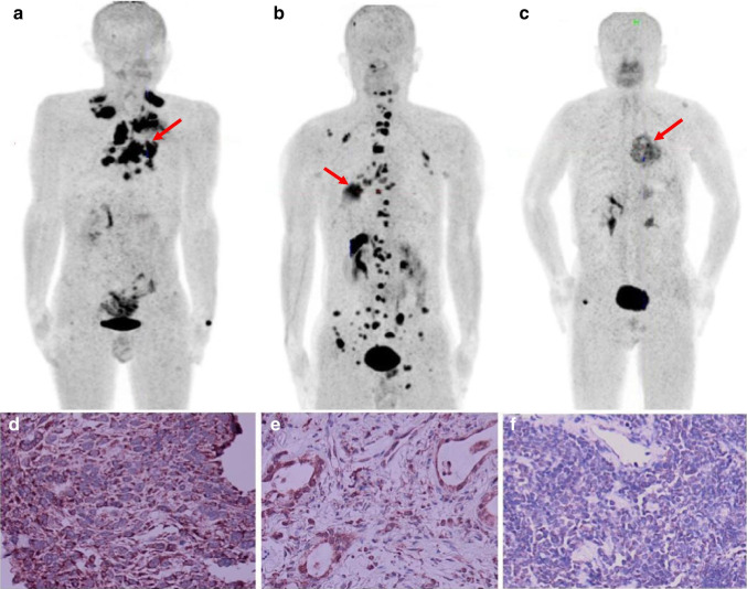
Heterogeneity is found in the tumor microenvironment among different pathological types of tumors. Radionuclide-labeled fibroblast-activation-protein inhibitor (FAPI), as an important tracer for non-invasive imaging of the tumor microenvironment, can be used to evaluate the expression of FAP in cancer-associated fibroblasts, macrophages, and tumor cells. Our aim was to explore the ability of [F]AlF-NOTA-FAPI-04 positron emission tomography (PET)/computed tomography (CT) to distinguish different types of lung cancer by evaluating the uptake of this tracer in primary and metastatic lesions.

METHODS

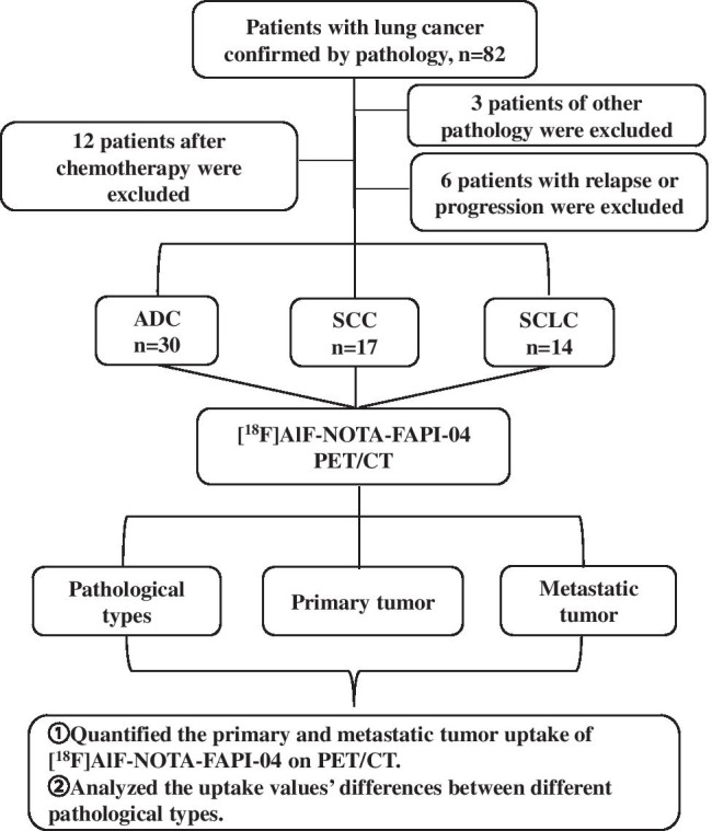
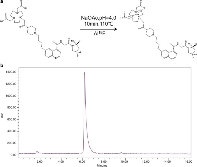
We prospectively enrolled 61 patients with histopathologically proven primary lung cancer with metastases. PET/CT scanning was performed before any antitumor therapy and 1 h after injection of 235.10 ± 3.89 MBq of [F]AlF-NOTA-FAPI-04. Maximum standard uptake values (SUV) were calculated for comparison among primary and metastatic lesions. Immunohistochemical staining for FAP was performed on tumor specimens.

RESULTS

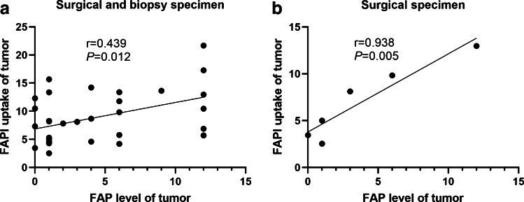
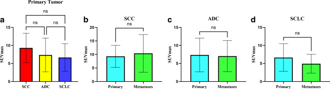
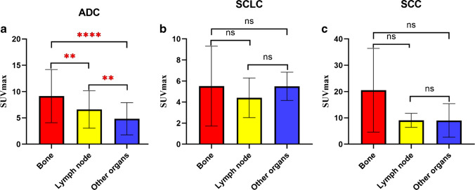
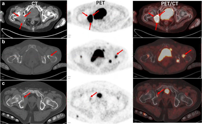
Sixty-one patients with adenocarcinoma (ADC, n = 30), squamous cell carcinoma (SCC, n = 17), and small cell lung cancer (SCLC, n = 14) were enrolled in this study, and 61 primary tumors and 199 metastases were evaluated. No difference in [F]AlF-NOTA-FAPI-04 uptake was observed among primary ADC, SCC, and SCLC tumors (P = 0.198). Additionally, no difference in uptake was found between primary and metastatic lesions of lung cancer with the same pathological type (P > 0.05). However, uptake did differ among metastases of differing pathological types (P < 0.001). The SUV of metastatic lymph nodes was highest for SCC, followed by ADC and then SCLC (P < 0.001). The SUV of bone metastases also was highest for SCC, followed by ADC and SCLC (P < 0.05), but no difference was observed between ADC and SCLC. The SUV of metastases in other organs was higher in SCC cases than in ADC cases but did not differ between SCC and SCLC or ADC and SCLC. Bone metastases exhibited higher uptake than those of lymph nodes and other organs in SCC and ADC (P < 0.05) but not in SCLC. Positive correlations were found between FAPI uptake and FAP expression in surgical plus biopsy specimens (r = 0.439, P = 0.012) and surgical specimens (r = 0.938, P = 0.005).

CONCLUSION

[F]AlF-NOTA-FAPI-04 PET/CT imaging revealed differences in FAP expression in metastases of lung cancer, with the highest expression specifically in bone metastases, and thus, may be valuable for distinguishing different pathological types of lung cancer.

摘要

目的

不同病理类型的肿瘤之间存在肿瘤微环境的异质性。放射性核素标记的成纤维细胞激活蛋白抑制剂(FAPI)作为肿瘤微环境非侵入性成像的重要示踪剂,可用于评估癌症相关成纤维细胞、巨噬细胞和肿瘤细胞中 FAP 的表达。我们的目的是通过评估原发性和转移性病变中该示踪剂的摄取,探讨[F]AlF-NOTA-FAPI-04 正电子发射断层扫描(PET)/计算机断层扫描(CT)区分不同类型肺癌的能力。

方法

我们前瞻性纳入了 61 例经组织病理学证实的原发性肺癌伴转移患者。在任何抗肿瘤治疗前和注射[F]AlF-NOTA-FAPI-04 235.10±3.89MBq 1 小时后进行 PET/CT 扫描。比较原发性和转移性病变的最大标准摄取值(SUV)。对肿瘤标本进行 FAP 免疫组织化学染色。

结果

本研究纳入了 61 例腺癌(ADC,n=30)、鳞状细胞癌(SCC,n=17)和小细胞肺癌(SCLC,n=14)患者,共评估了 61 个原发性肿瘤和 199 个转移灶。ADC、SCC 和 SCLC 原发性肿瘤之间[F]AlF-NOTA-FAPI-04 摄取无差异(P=0.198)。此外,同一病理类型的肺癌原发性和转移性病变之间摄取无差异(P>0.05)。然而,不同病理类型的转移灶之间摄取有差异(P<0.001)。SCC 患者的转移性淋巴结 SUV 最高,其次是 ADC 和 SCLC(P<0.001)。SCC 患者的骨转移 SUV 也最高,其次是 ADC 和 SCLC(P<0.05),但 ADC 和 SCLC 之间无差异。SCC 患者其他器官转移的 SUV 高于 ADC 患者,但 SCC 与 SCLC 或 ADC 与 SCLC 之间无差异。SCC 患者的骨转移摄取高于淋巴结和其他器官转移(P<0.05),但 SCLC 患者无差异。手术加活检标本(r=0.439,P=0.012)和手术标本(r=0.938,P=0.005)中 FAPI 摄取与 FAP 表达呈正相关。

结论

[F]AlF-NOTA-FAPI-04 PET/CT 成像显示肺癌转移中 FAP 表达存在差异,在骨转移中表达最高,因此可能对区分不同类型的肺癌有价值。

https://cdn.ncbi.nlm.nih.gov/pmc/blobs/62e9/8940861/0226ad836026/259_2021_5638_Fig8_HTML.jpg
https://cdn.ncbi.nlm.nih.gov/pmc/blobs/62e9/8940861/1915cc4d1bbe/259_2021_5638_Fig1_HTML.jpg
https://cdn.ncbi.nlm.nih.gov/pmc/blobs/62e9/8940861/ae3b74f02227/259_2021_5638_Fig2_HTML.jpg
https://cdn.ncbi.nlm.nih.gov/pmc/blobs/62e9/8940861/c3a463ca31d3/259_2021_5638_Fig3_HTML.jpg
https://cdn.ncbi.nlm.nih.gov/pmc/blobs/62e9/8940861/5f5c8b61d291/259_2021_5638_Fig4_HTML.jpg
https://cdn.ncbi.nlm.nih.gov/pmc/blobs/62e9/8940861/725840042295/259_2021_5638_Fig5_HTML.jpg
https://cdn.ncbi.nlm.nih.gov/pmc/blobs/62e9/8940861/96f85de04253/259_2021_5638_Fig6_HTML.jpg
https://cdn.ncbi.nlm.nih.gov/pmc/blobs/62e9/8940861/666cccfac077/259_2021_5638_Fig7_HTML.jpg
https://cdn.ncbi.nlm.nih.gov/pmc/blobs/62e9/8940861/0226ad836026/259_2021_5638_Fig8_HTML.jpg

相似文献

1
[F]AlF-NOTA-FAPI-04 PET/CT uptake in metastatic lesions on PET/CT imaging might distinguish different pathological types of lung cancer.[F]氟代阿尔伐斯汀环丙基非肽类 FAPI-04 正电子发射断层扫描/计算机断层扫描(PET/CT)摄取在转移灶的 PET/CT 成像上可能有助于鉴别不同病理类型的肺癌。
Eur J Nucl Med Mol Imaging. 2022 Apr;49(5):1671-1681. doi: 10.1007/s00259-021-05638-z. Epub 2021 Dec 6.
2
Value of [F]AlF-NOTA-FAPI-04 PET/CT for differential diagnosis of malignant and various inflammatory lung lesions: comparison with [F]FDG PET/CT.^18^F-AlF-NOTA-FAPI-04 PET/CT 对良恶性及各种肺部炎性病变的鉴别诊断价值:与^18^F-FDG PET/CT 的比较。
Eur Radiol. 2024 Mar;34(3):1948-1959. doi: 10.1007/s00330-023-10208-y. Epub 2023 Sep 5.
3
[F]AlF-NOTA-FAPI-04: FAP-targeting specificity, biodistribution, and PET/CT imaging of various cancers.[F]AlF-NOTA-FAPI-04:针对各种癌症的 FAP 靶向特异性、生物分布和 PET/CT 成像。
Eur J Nucl Med Mol Imaging. 2022 Jul;49(8):2761-2773. doi: 10.1007/s00259-022-05758-0. Epub 2022 Mar 9.
4
Preclinical evaluation and pilot clinical study of [F]AlF-NOTA-FAPI-04 for PET imaging of rheumatoid arthritis.[F]AlF-NOTA-FAPI-04 的临床前评估和初步临床研究用于类风湿关节炎的 PET 成像。
Eur J Nucl Med Mol Imaging. 2022 Oct;49(12):4025-4036. doi: 10.1007/s00259-022-05836-3. Epub 2022 Jun 18.
5
PET Imaging of Fibroblast Activation Protein in Various Types of Cancer Using Ga-FAP-2286: Comparison with F-FDG and Ga-FAPI-46 in a Single-Center, Prospective Study.使用 Ga-FAP-2286 对多种癌症中的成纤维细胞激活蛋白进行 PET 成像:在单中心前瞻性研究中与 F-FDG 和 Ga-FAPI-46 的比较。
J Nucl Med. 2023 Mar;64(3):386-394. doi: 10.2967/jnumed.122.264544. Epub 2022 Sep 2.
6
[F] AlF‑NOTA‑FAPI‑04 PET/CT as a promising tool for imaging fibroblast activation protein in gastrointestinal system cancers: a prospective investigation of comparative analysis with F-FDG.[F] AlF‑NOTA‑FAPI‑04 PET/CT 作为一种有前途的成像工具,用于胃肠道癌症中的成纤维细胞激活蛋白:与 F-FDG 比较分析的前瞻性研究。
Eur J Nucl Med Mol Imaging. 2023 Nov;50(13):4051-4063. doi: 10.1007/s00259-023-06351-9. Epub 2023 Aug 5.
7
[F]FAPI-42 PET/CT versus [F]FDG PET/CT for imaging of recurrent or metastatic gastrointestinal stromal tumors.FAPI-42 PET/CT 与 [F]FDG PET/CT 对比用于复发或转移性胃肠道间质瘤的成像。
Eur J Nucl Med Mol Imaging. 2022 Dec;50(1):194-204. doi: 10.1007/s00259-022-05955-x. Epub 2022 Aug 30.
8
Improved cancer detection in Waldeyer's tonsillar ring by Ga-FAPI PET/CT imaging.Ga-FAPI PET/CT 成像提高了咽扁桃体环的癌症检测能力。
Eur J Nucl Med Mol Imaging. 2021 Apr;48(4):1178-1187. doi: 10.1007/s00259-020-05055-8. Epub 2020 Oct 15.
9
Comparison of 18 F-FDG PET/CT and 68 Ga-FAPI-04 PET/CT in patients with non-small cell lung cancer.18 F-FDG PET/CT 与 68 Ga-FAPI-04 PET/CT 在非小细胞肺癌患者中的比较。
Nucl Med Commun. 2022 Oct 1;43(10):1084-1091. doi: 10.1097/MNM.0000000000001607. Epub 2022 Aug 17.
10
Visualization of Activated Fibroblasts in Heart Failure with Preserved Ejection Fraction with [F]AlF-NOTA-FAPI-04 PET/CT Imaging.[F]AlF-NOTA-FAPI-04 PET/CT 显像在射血分数保留心力衰竭中活化成纤维细胞的可视化。
Mol Pharm. 2023 May 1;20(5):2634-2641. doi: 10.1021/acs.molpharmaceut.3c00075. Epub 2023 Apr 12.

引用本文的文献

1
Clinical applications of fibroblast activation protein inhibitor positron emission tomography (FAPI-PET).成纤维细胞活化蛋白抑制剂正电子发射断层扫描(FAPI-PET)的临床应用
Npj Imaging. 2024 Nov 13;2(1):48. doi: 10.1038/s44303-024-00053-z.
2
Detecting radiation esophagitis using F-FAPI-04 PET/CT in patients with LA-ESCC treated with concurrent chemoradiotherapy.使用F-FAPI-04 PET/CT检测接受同步放化疗的LA-ESCC患者的放射性食管炎。
BMC Cancer. 2025 May 12;25(1):854. doi: 10.1186/s12885-025-14236-3.
3
Noninvasive diagnosis and classification of kidney transplantation rejection by F-FAPI-04 PET/CT.

本文引用的文献

1
Screening and identification of key biomarkers in lung squamous cell carcinoma by bioinformatics analysis.通过生物信息学分析筛选和鉴定肺鳞状细胞癌中的关键生物标志物
Oncol Lett. 2019 Nov;18(5):5185-5196. doi: 10.3892/ol.2019.10873. Epub 2019 Sep 16.
2
Prognostic effects of osteoclast inhibitors in extensive stage small cell lung cancer patients with bone metastases.破骨细胞抑制剂对广泛期小细胞肺癌骨转移患者的预后影响。
Mol Clin Oncol. 2018 Nov;9(5):561-565. doi: 10.3892/mco.2018.1710. Epub 2018 Sep 4.
通过F-FAPI-04 PET/CT对肾移植排斥反应进行无创诊断和分类
Eur J Nucl Med Mol Imaging. 2025 May 8. doi: 10.1007/s00259-025-07307-x.
4
[F]FAPI- 04 PET/CT for pathologic response assessment in pancreatic cancer patients with systemic treatment.[F]FAPI-04正电子发射断层显像/计算机断层扫描用于接受全身治疗的胰腺癌患者的病理反应评估
Eur J Nucl Med Mol Imaging. 2025 Apr 16. doi: 10.1007/s00259-025-07271-6.
5
Relationship between PD-L1 expression and [F]FAPI versus [F]FDG uptake on PET/CT in lung cancer.肺癌患者PET/CT上PD-L1表达与[F]FAPI及[F]FDG摄取之间的关系
Eur J Nucl Med Mol Imaging. 2025 Mar 21. doi: 10.1007/s00259-025-07201-6.
6
Comparison of the diagnostic accuracy between F-FAPI-04 PET/CT and F-FDG PET/CT in the clinical stage IA of lung adenocarcinoma.F-FAPI-04 PET/CT与F-FDG PET/CT在肺腺癌临床IA期诊断准确性的比较。
J Thorac Dis. 2025 Feb 28;17(2):661-675. doi: 10.21037/jtd-24-1658. Epub 2025 Feb 27.
7
Value of [F]AlF-NOTA-FAPI-04 PET/CT for predicting pathological response and survival in patients with locally advanced pancreatic ductal adenocarcinoma receiving neoadjuvant chemotherapy.[F]AlF-NOTA-FAPI-04 PET/CT在预测接受新辅助化疗的局部晚期胰腺导管腺癌患者病理反应和生存情况中的价值
Eur J Nucl Med Mol Imaging. 2025 May;52(6):2118-2131. doi: 10.1007/s00259-025-07084-7. Epub 2025 Jan 17.
8
[F]F-FAPI-42 PET dynamic imaging characteristics and multiparametric quantification of lung cancer: an exploratory study using uEXPLORER PET/CT.[F]F-FAPI-42 PET对肺癌的动态成像特征及多参数定量分析:一项使用uEXPLORER PET/CT的探索性研究
Eur J Nucl Med Mol Imaging. 2025 Apr;52(5):1685-1694. doi: 10.1007/s00259-024-07064-3. Epub 2025 Jan 6.
9
Evaluating the diagnostic value of F-FAPI-04 PET/CT in various malignant tumors: a head-to-head comparison with F-FDG PET/CT.评估F-FAPI-04 PET/CT在各种恶性肿瘤中的诊断价值:与F-FDG PET/CT的直接比较。
Jpn J Radiol. 2025 May;43(5):852-863. doi: 10.1007/s11604-024-01714-0. Epub 2024 Dec 4.
10
Comparative assessment of the diagnostic efficacy of [F]AlF-NOTA-FAPI-04 and [F]FDG PET/CT imaging for detecting postoperative recurrence in gastric cancer patients: a pilot study.[F]AlF-NOTA-FAPI-04与[F]FDG PET/CT成像检测胃癌患者术后复发的诊断效能比较评估:一项初步研究。
Front Oncol. 2024 Sep 11;14:1427649. doi: 10.3389/fonc.2024.1427649. eCollection 2024.