Laboratory of Neuroimaging, National Institute on Alcohol Abuse and Alcoholism, National Institutes of Health, Bethesda, Maryland, USA.
Department of Diagnostic Radiology, Yale University School of Medicine, New Haven, Connecticut, USA.
Hum Brain Mapp. 2022 Mar;43(4):1419-1430. doi: 10.1002/hbm.25733. Epub 2021 Dec 7.
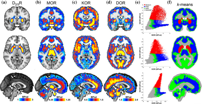
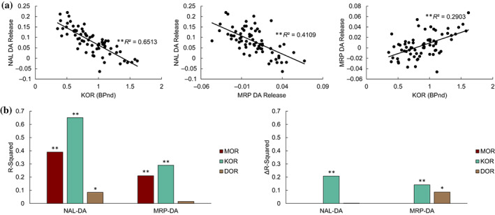
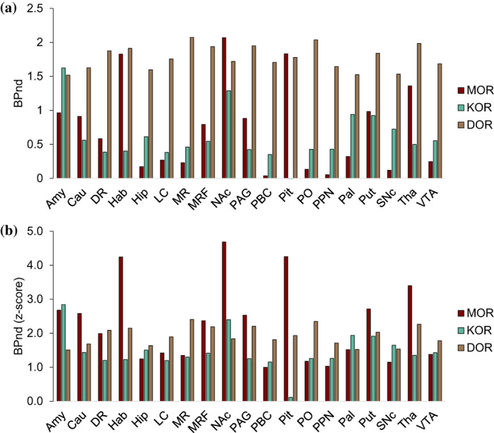
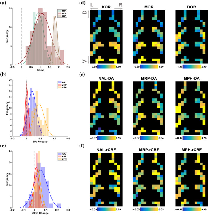
Opioid receptors are expressed throughout the brain and play a major role in regulating striatal dopamine (DA) release. Clinical studies have shown that naloxone (NAL, a nonspecific opioid antagonist) in individuals with opioid use disorder and morphine (MRP, a nonspecific opioid agonist) in healthy controls, resulted in DA release in the dorsal and ventral striatum, respectively. It is not known whether the underlying patterns of striatal DA release are associated with the striatal distribution of opioid receptors. We leveraged previously published PET datasets (collected in independent cohorts) to study the brain-wide distribution of opioid receptors and to compare striatal opioid receptor availability with striatal DA release patterns. We identified three major gray matter segments based on availability maps of DA and opioid receptors: striatum, and primary and secondary opioid segments with high and intermediate opioid receptor availability, respectively. Patterns of DA release induced by NAL and MRP were inversely associated and correlated with kappa (NAL: r(68) = -0.81, MRP: r(68) = 0.54), and mu (NAL: r(68) = -0.62, MRP: r(68) = 0.46) opioid receptor availability. Kappa opioid receptor availability accounted for a unique part of variance in NAL- and MRP-DA release patterns (ΔR >0.14, p <.0001). In sum, distributions of opioid receptors distinguished major cortical and subcortical regions. Patterns of NAL- and MRP-induced DA release had inverse associations with striatal opioid receptor availability. Our approach provides a pattern-based characterization of drug-induced DA targets and is relevant for modeling the role of opioid receptors in modulating striatal DA release.
阿片受体广泛表达于大脑中,在调节纹状体多巴胺(DA)释放方面发挥着主要作用。临床研究表明,纳洛酮(NAL,一种非特异性阿片拮抗剂)在阿片类药物使用障碍患者中,以及吗啡(MRP,一种非特异性阿片激动剂)在健康对照者中,分别导致背侧和腹侧纹状体的 DA 释放。目前尚不清楚纹状体 DA 释放的潜在模式是否与阿片受体在纹状体中的分布有关。我们利用先前发表的 PET 数据集(在独立队列中收集)来研究阿片受体在大脑中的广泛分布,并比较纹状体阿片受体的可用性与纹状体 DA 释放模式。我们根据 DA 和阿片受体的可用性图谱,确定了三个主要的灰质节段:纹状体,以及初级和次级阿片节段,它们分别具有高和中等阿片受体可用性。NAL 和 MRP 诱导的 DA 释放模式呈负相关,并与κ(NAL:r(68)= -0.81,MRP:r(68)= 0.54)和μ(NAL:r(68)= -0.62,MRP:r(68)= 0.46)阿片受体可用性相关。κ阿片受体可用性解释了 NAL 和 MRP-DA 释放模式中独特的方差部分(ΔR >0.14,p <.0001)。总之,阿片受体的分布区分了主要的皮质和皮质下区域。NAL 和 MRP 诱导的 DA 释放模式与纹状体阿片受体的可用性呈负相关。我们的方法提供了一种基于模式的药物诱导 DA 靶点特征描述,对于模拟阿片受体在调节纹状体 DA 释放中的作用具有重要意义。