Department of Neurology, Affiliated ZhongDa Hospital, School of Medicine, Research Institution of Neuropsychiatry, Southeast University, Nanjing, 210009, China.
Center of Interventional Radiology and Vascular Surgery, Department of Radiology, Affiliated Zhongda Hospital, Medical School, Southeast University, Nanjing, 210009, China.
J Neuroinflammation. 2021 Dec 7;18(1):283. doi: 10.1186/s12974-021-02333-6.
Inflammation is integral to the pathophysiology of ischemic stroke and a prime target for the development of new stroke therapies. The aim of the present study is to seek out the regulatory mechanism of circCDC14A in neuroinflammatory injury in tMCAO mice.
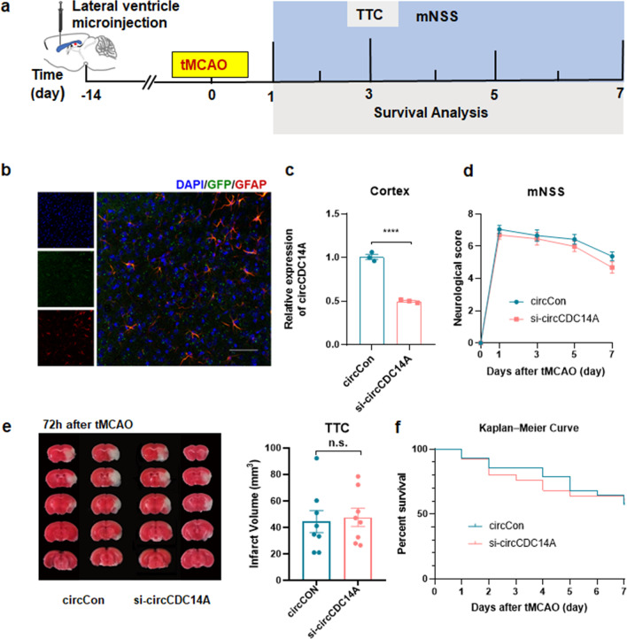
The expression level of circCDC14A in peri-infarct cortex and plasma of mice were detected by qPCR. The localization of circCDC14A in peripheral blood cells and peri-infarct cortex of tMCAO mice were explored by in situ hybridization and immunofluorescence colocalization staining. Lentivirus were microinjected into lateral ventricular of brain or injected into tail vein to interfere with the expression of circCDC14A, thus their effects on behavior, morphology, and molecular biology of tMCAO mice were analyzed.
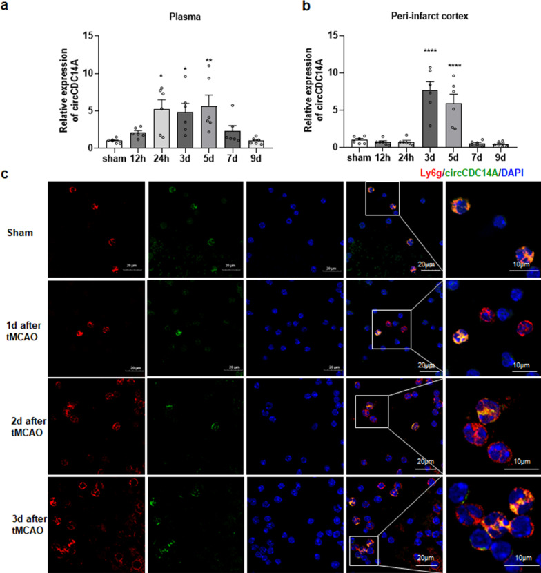
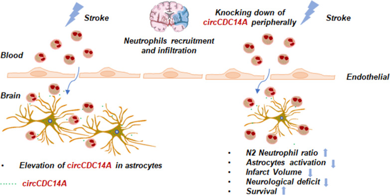
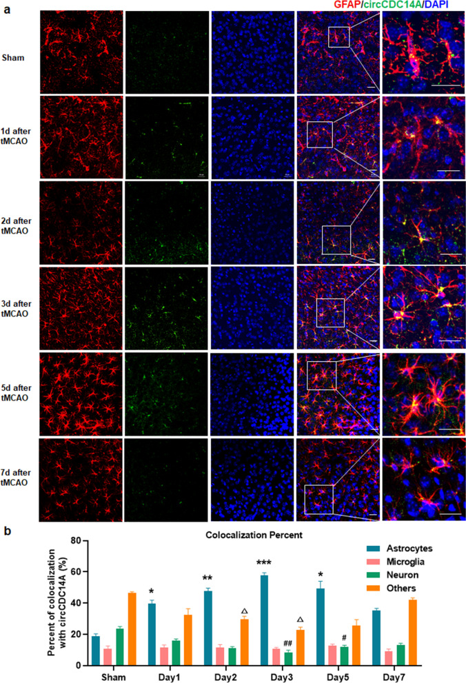
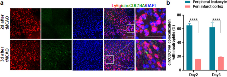
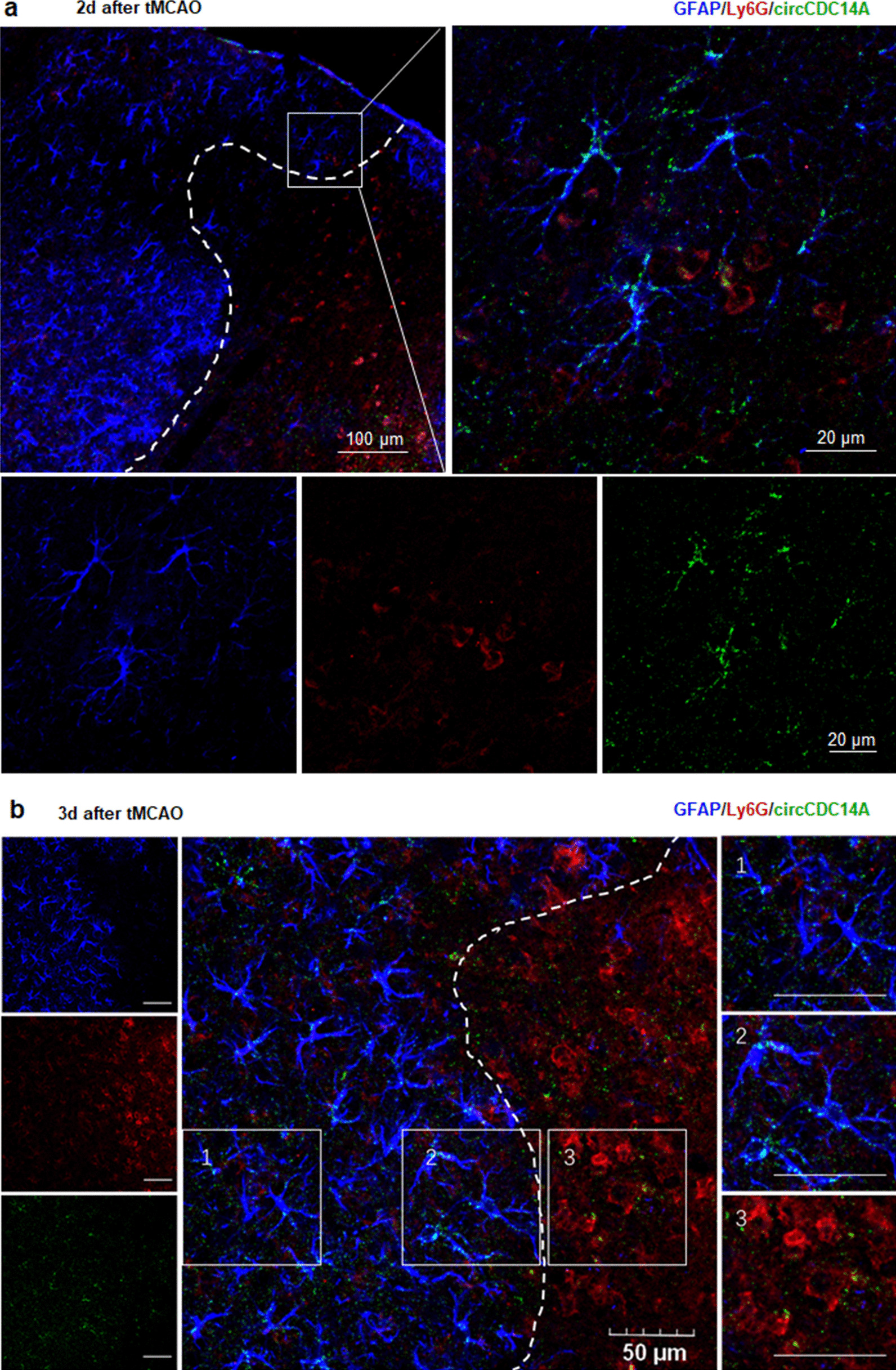
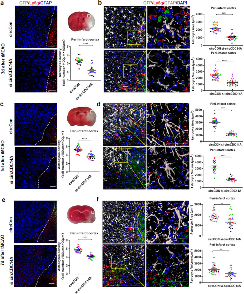
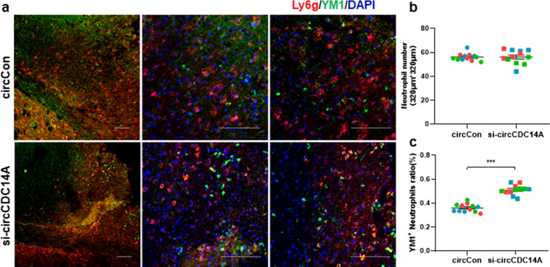
The expression of circCDC14A in plasma and peri-infarct cortex of tMCAO mice significantly increased, and circCDC14A was mainly localized in neutrophils peripherally while in astrocytes in peri-infarct cortex centrally. Tail vein injection of lentivirus to interfere with the expression of circCDC14A significantly reduced the infarct volume (P < 0.01) at 72 h after reperfusion and density of activated astrocytes in peri-infarct cortex at 3 days, 5 days and 7 days after tMCAO modeling (all P < 0.0001). Moreover, mNSS (P < 0.0001) and survival rate (P < 0.001) were significantly improved within 7 days in si-circCDC14A group compared to circCon group. Additionally, morphology analysis showed the volume and surface area of each activated astrocytes significantly decreased (P < 0.0001). Quantification analysis measured the percentage of N2 phenotype among infiltrated neutrophils in brain sections and found N2 ratio was significantly higher in si-circCDC14A group compared to circCon group (P < 0.001).
Knocking down the expression of circCDC14A in peripheral blood cells relieved astrocytes activation in peri-infarct cortex, thereby relieved brain damage in the acute phase of ischemic stroke.
炎症是缺血性中风病理生理学的重要组成部分,也是开发新中风治疗方法的主要靶点。本研究旨在寻找 circCDC14A 在 tMCAO 小鼠神经炎症损伤中的调控机制。
通过 qPCR 检测小鼠梗死周边皮质和血浆中 circCDC14A 的表达水平。通过原位杂交和免疫荧光共定位染色探索 tMCAO 小鼠外周血单个核细胞和梗死周边皮质中 circCDC14A 的定位。通过脑侧脑室微注射或尾静脉注射慢病毒来干扰 circCDC14A 的表达,从而分析其对 tMCAO 小鼠行为、形态和分子生物学的影响。
tMCAO 小鼠血浆和梗死周边皮质中 circCDC14A 的表达显著增加,circCDC14A 主要定位于外周血中的中性粒细胞,而在梗死周边皮质中主要定位于星形胶质细胞。尾静脉注射慢病毒干扰 circCDC14A 的表达,可显著减少再灌注后 72 小时的梗死体积(P<0.01),并降低 tMCAO 模型后 3、5 和 7 天梗死周边皮质中激活星形胶质细胞的密度(均 P<0.0001)。此外,在 7 天内,与 circCon 组相比,si-circCDC14A 组的 mNSS(P<0.0001)和存活率(P<0.001)均显著提高。此外,形态学分析表明,每个激活的星形胶质细胞的体积和表面积均显著减小(P<0.0001)。对脑切片中浸润的中性粒细胞的 N2 表型的百分比进行定量分析,发现 si-circCDC14A 组中的 N2 比值明显高于 circCon 组(P<0.001)。
敲低外周血单个核细胞中的 circCDC14A 表达可减轻梗死周边皮质中星形胶质细胞的激活,从而减轻缺血性中风急性期的脑损伤。