Wang Min, Liu Hai-Feng, Zhang Yan-Zhen-Zi, Zou Zhi-Qing, Wu Zhou-Quan
Department of Anesthesiology, Changzhou Second People's Hospital Affiliated to Nanjing Medical University, Changzhou 213003, Jiangsu Province, China.
Department of Radiology, Third Affiliated Hospital of Soochow University and Changzhou First People's Hospital, Changzhou 213003, Jiangsu Province, China.
World J Clin Cases. 2021 Nov 16;9(32):9948-9953. doi: 10.12998/wjcc.v9.i32.9948.
Hepatic hemolymphangioma is an extremely rare benign congenital malformation composed of cystically dilated lymphatic and blood vessels, and they have nonspecific clinical symptoms and laboratory results. In this study, hepatic hemolymphangioma with multiple hemangiomas in an elderly woman was initially reported and analyzed.
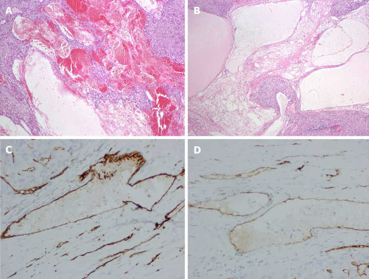
A 61-year-old female patient, with a history of hysterectomy and bilateral adnexectomy, was referred to the hepatobiliary surgery department with the complaint of multiple hepatic hemangiomas that had been diagnosed 2 years prior in a preoperative contrast-enhanced computed tomography (CECT) examination. Upon entering our hospital, no abnormal physical examination and laboratory data were found. The latest CECT revealed a new 7.0 cm × 6.2 cm cystic-solid lesion with multiple internal divisions in segment II of the liver, with delayed CECT enhancement characteristics that presented as solid parts with internal division. On the positron emission tomography (PET)/CT, no significant uptake of F-fluorodeoxyglucse was observed. Finally, hepatic hemolymphangioma was confirmed based on the pathological and immunohistochemical results after surgery. At 1-year follow-up, her posthepatectomy evaluation was uneventful, and she had recovered full activity. In addition, no postoperative recurrent or residual lesion was found on CECT imaging.
Hepatic hemolymphangioma with multiple hemangiomas was reported and observed by CECT and PET/CT imaging.
肝血管淋巴管瘤是一种极其罕见的良性先天性畸形,由囊性扩张的淋巴管和血管组成,其临床症状和实验室检查结果无特异性。本研究首次报道并分析了一名老年女性患有多发血管瘤的肝血管淋巴管瘤病例。
一名61岁女性患者,有子宫切除和双侧附件切除病史,因2年前术前增强CT检查诊断为多发肝血管瘤而转诊至肝胆外科。入院时,体格检查和实验室检查均未发现异常。最新的增强CT显示肝脏Ⅱ段有一个新的7.0 cm×6.2 cm囊实性病变,内部有多个分隔,增强CT延迟强化表现为内部有分隔的实性部分。在正电子发射断层扫描(PET)/CT上,未观察到氟脱氧葡萄糖有明显摄取。最终,根据术后病理和免疫组化结果确诊为肝血管淋巴管瘤。随访1年,肝切除术后评估无异常,患者已恢复全部活动。此外,增强CT成像未发现术后复发或残留病变。
报道了多发血管瘤的肝血管淋巴管瘤病例,并通过增强CT和PET/CT成像进行了观察。