Braun Clarissa, Katholnig Karl, Kaltenecker Christopher, Linke Monika, Sukhbaatar Nyamdelger, Hengstschläger Markus, Weichhart Thomas
Center of Pathobiochemistry and Genetics, Institute of Medical Genetics, Medical University of Vienna, Vienna, Austria.
Clinical Division of Endocrinology and Metabolism, Department of Internal Medicine III, Medical University of Vienna, Vienna, Austria.
Cell Stress. 2021 Nov 23;5(12):176-182. doi: 10.15698/cst2021.12.260. eCollection 2021 Dec.
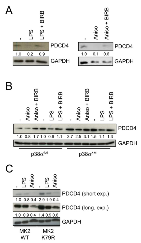
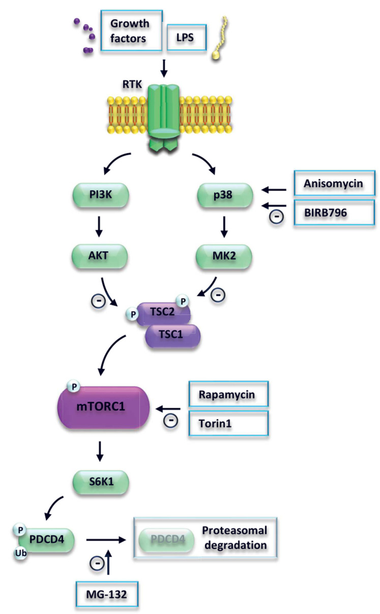
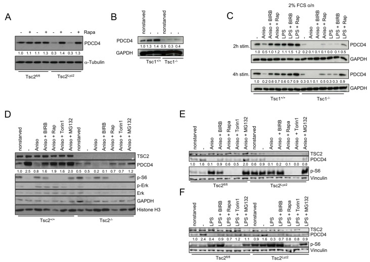
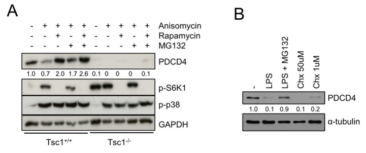
Programmed cell death protein 4 (PDCD4) exerts critical functions as tumor suppressor and in immune cells to regulate inflammatory processes. The phosphoinositide 3-kinase (PI3K) promotes degradation of PDCD4 via mammalian target of rapamycin complex 1 (mTORC1). However, additional pathways that may regulate PDCD4 expression are largely ill-defined. In this study, we have found that activation of the mitogen-activated protein kinase p38 promoted degradation of PDCD4 in macrophages and fibroblasts. Mechanistically, we identified a pathway from p38 and its substrate MAP kinase-activated protein kinase 2 (MK2) to the tuberous sclerosis complex (TSC) to regulate mTORC1-dependent degradation of PDCD4. Moreover, we provide evidence that TSC1 and TSC2 regulate PDCD4 expression via an additional mechanism independent of mTORC1. These novel data extend our knowledge of how PDCD4 expression is regulated by stress- and nutrient-sensing pathways.
程序性细胞死亡蛋白4(PDCD4)作为肿瘤抑制因子以及在免疫细胞中发挥关键功能,以调节炎症过程。磷酸肌醇3激酶(PI3K)通过雷帕霉素哺乳动物靶蛋白复合物1(mTORC1)促进PDCD4的降解。然而,可能调节PDCD4表达的其他途径在很大程度上尚不明确。在本研究中,我们发现丝裂原活化蛋白激酶p38的激活促进了巨噬细胞和成纤维细胞中PDCD4的降解。从机制上来说,我们确定了一条从p38及其底物丝裂原活化蛋白激酶激活的蛋白激酶2(MK2)到结节性硬化复合物(TSC)的途径,以调节mTORC1依赖的PDCD4降解。此外,我们提供证据表明TSC1和TSC2通过一种独立于mTORC1的额外机制调节PDCD4的表达。这些新数据扩展了我们对PDCD4表达如何受应激和营养感知途径调节的认识。