Department of Radiology, Azienda USL - IRCCS di Reggio Emilia, Reggio Emilia, Italy.
Unit of Rheumatology, Azienda USL - IRCCS di Reggio Emilia, Reggio Emilia, Italy.
RMD Open. 2022 Jan;8(1). doi: 10.1136/rmdopen-2021-001977.
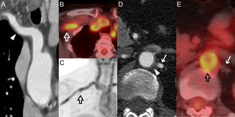
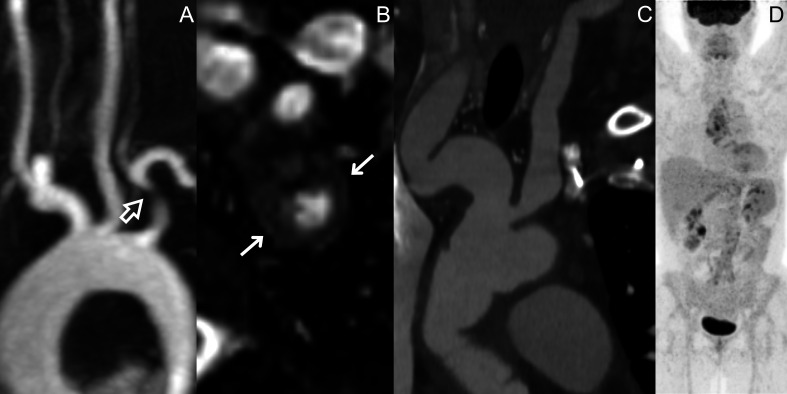
The aim was to identify any association between imaging signs of vessel wall inflammation (positron emission tomography-CT (PET-CT) score and CT/MR wall thickening) and synchronous and subsequent vascular damage (stenoses/dilations) in patients with large vessel vasculitis (LVV).
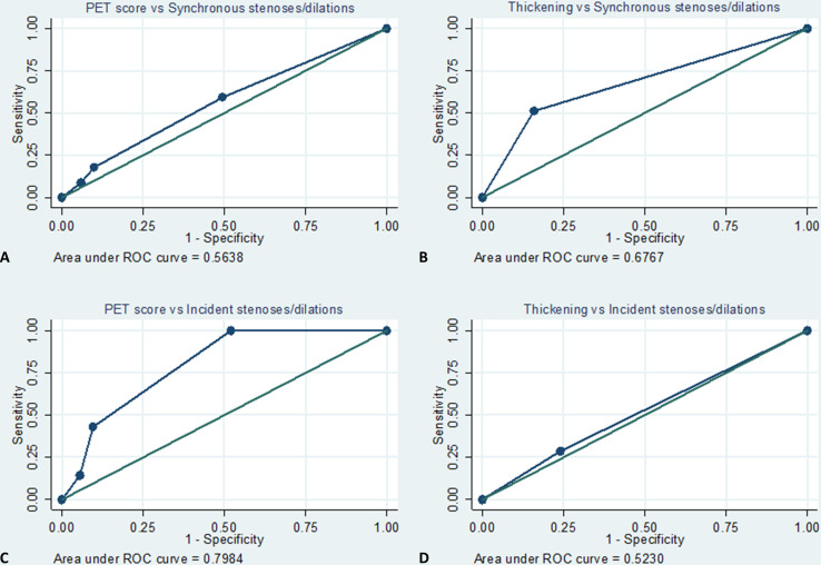
Consecutive patients with LVV referred to a tertiary centre in 2007-2020 with baseline PET-CT and morphological imaging (CT/MR angiography) performed within 3 months were included. All available PET-CT and CT/MR scans were reviewed to assess PET-CT uptake (4-point semi-quantitative score), wall thickening, stenoses and dilations for 15 vascular segments. The associations of baseline PET score and CT/MR wall thickening with synchronous and incident stenoses/dilations at CT/MR performed 6-30 months from baseline were evaluated in per-segment and per-patient analyses. Respective areas under the receiver operating characteristic curve (AUC) were calculated.
We included 100 patients with LVV (median age: 48 years, 22% males). Baseline PET score and wall thickening were strongly associated (Cuzick non-parametric test for trend across order groups (NPtrend) <0.001). The association with synchronous stenoses/dilations was weak for PET score (NPtrend=0.01) and strong for wall thickening (p<0.001). In per-patient analyses, sensitivity/specificity for ≥1 synchronous stenoses/dilations were 44%/67% for PET score ≥2 and 66.7%/60.5% for wall thickening. Subsequent CTs/MRs were available in 28 patients, with seven incident stenoses/dilations. Baseline PET score was strongly associated with incident stenoses/dilations (p=0.001), while baseline wall thickening was not (p=0.708), with AUCs for incident stenoses/dilations of 0.80 for PET score and 0.52 for wall thickening.
PET score and wall thickening are strongly associated, but only baseline PET score is a good predictor of incident vessel wall damage in LVV.
本研究旨在确定血管壁炎症的影像学征象(正电子发射断层扫描计算机断层扫描(PET-CT)评分和 CT/MR 管壁增厚)与大血管血管炎(LVV)患者的同步和随后的血管损伤(狭窄/扩张)之间是否存在关联。
本研究纳入了 2007 年至 2020 年期间连续就诊于三级医疗中心的 LVV 患者,基线 PET-CT 和形态学成像(CT/MR 血管造影)在 3 个月内完成。对所有可获得的 PET-CT 和 CT/MR 扫描进行回顾性分析,以评估 15 个血管节段的 PET-CT 摄取(4 分半定量评分)、管壁增厚、狭窄和扩张。采用分段和整体患者分析评估基线 PET 评分和 CT/MR 管壁增厚与基线后 6-30 个月 CT/MR 检查时同步和新发狭窄/扩张的相关性。计算各自的受试者工作特征曲线(ROC)下面积(AUC)。
本研究共纳入了 100 例 LVV 患者(中位年龄为 48 岁,22%为男性)。基线 PET 评分和管壁增厚呈强相关(Cuzick 非参数趋势检验(NPtrend)<0.001)。PET 评分与同步狭窄/扩张的相关性较弱(NPtrend=0.01),而管壁增厚的相关性较强(p<0.001)。在整体患者分析中,对于≥1 个同步狭窄/扩张,PET 评分≥2 的敏感性/特异性为 44%/67%,而管壁增厚的敏感性/特异性为 66.7%/60.5%。28 例患者随后进行了 CT/MR 检查,发现了 7 例新发狭窄/扩张。基线 PET 评分与新发狭窄/扩张显著相关(p=0.001),而基线管壁增厚与新发狭窄/扩张无显著相关(p=0.708),PET 评分预测新发狭窄/扩张的 AUC 为 0.80,而管壁增厚的 AUC 为 0.52。
PET 评分和管壁增厚密切相关,但只有基线 PET 评分是 LVV 患者发生血管壁损伤的良好预测指标。