AP-HP, Sorbonne Université, Hôpital Saint-Antoine, Service de Médecine Interne and Inflammation-Immunopathology-Biotherapy Department (DHU i2B), F-75012, Paris, France.
IMIV, CEA, INSERM, Université Paris Sud, CNRS, Université Paris Saclay, Orsay, France.
Sci Rep. 2019 Aug 27;9(1):12388. doi: 10.1038/s41598-019-48709-w.
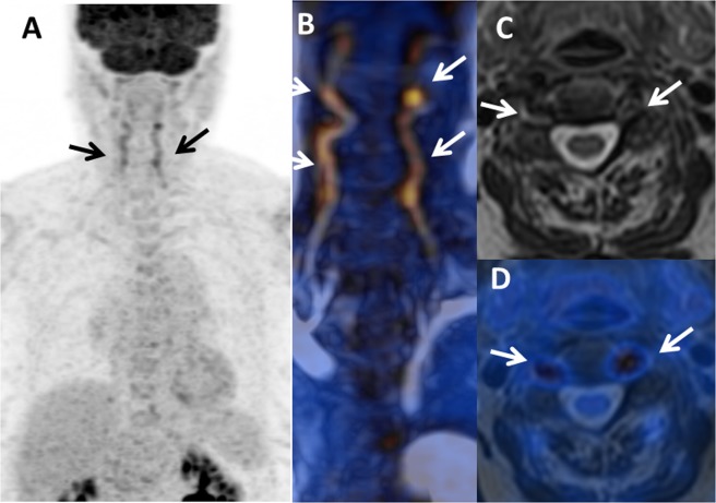
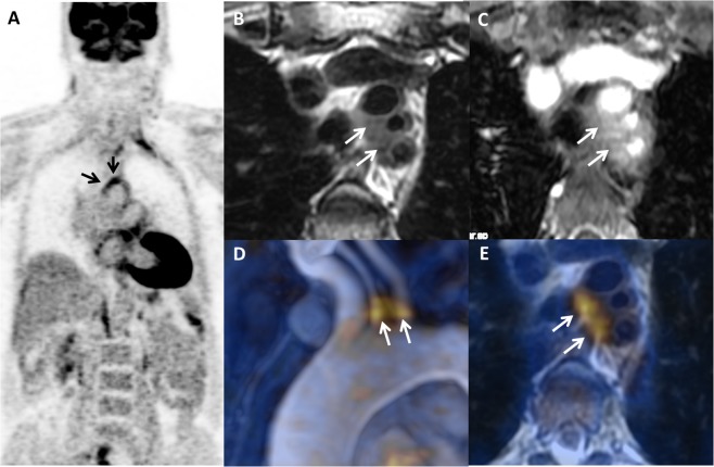
Diagnosis of large vessel vasculitis (LVV) and evaluation of its inflammatory activity can be challenging. Our aim was to investigate the value of hybrid positron-emission tomography/magnetic resonance imaging (PET/MRI) in LVV. All consecutive patients with LVV from the Department of Internal Medicine who underwent PET/MRI were included. Three PET/MRI patterns were defined: (i) "inflammatory," with positive PET (>liver uptake) and abnormal MRI (stenosis and/or wall thickening); (ii) "fibrous", negative PET (≤liver uptake) and abnormal MRI; and (iii) "normal". Thirteen patients (10 female; median age: 67-years [range: 23-87]) underwent 18 PET/MRI scans. PET/MRI was performed at diagnosis (n = 4), at relapse (n = 7), or during remission (n = 7). Among the 18 scans, eight (44%) showed an inflammatory pattern and three (17%) a fibrous pattern; the other seven were normal. The distribution of the three patterns did not differ between patients with Takayasu arteritis (TA, n = 10 scans) and those with giant cell arteritis (GCA, n = 8 scans). PET/MRI findings were normal in 2/10 (20%) TA scans vs. 5/8 (62%) GCA scans (p = 0.3). Median SUV was 4.7 [2.1-8.6] vs. 2 [1.8-2.6] in patients with active disease vs. remission, respectively (p = 0.003). PET/MRI is a new hybrid imaging modality allowing comprehensive and multimodal analysis of vascular wall inflammation and the vascular lumen. This technique offers promising perspectives for the diagnosis and monitoring of LVV.
大动脉炎(LVV)的诊断和其炎症活动的评估具有一定挑战性。我们旨在研究正电子发射断层扫描/磁共振成像(PET/MRI)在 LVV 中的价值。所有来自内科系并接受 PET/MRI 的 LVV 连续患者均纳入研究。定义了三种 PET/MRI 模式:(i)“炎症”,阳性 PET(>肝脏摄取)和异常 MRI(狭窄和/或壁增厚);(ii)“纤维”,阴性 PET(≤肝脏摄取)和异常 MRI;以及(iii)“正常”。13 名患者(10 名女性;中位年龄:67 岁[范围:23-87])接受了 18 次 PET/MRI 扫描。PET/MRI 在诊断时(n=4)、复发时(n=7)或缓解期(n=7)进行。在 18 次扫描中,8 次(44%)显示炎症模式,3 次(17%)显示纤维模式;其他 7 次正常。三种模式的分布在 Takayasu 动脉炎(TA,n=10 次扫描)和巨细胞动脉炎(GCA,n=8 次扫描)患者中无差异。2/10(20%)TA 扫描的 PET/MRI 结果正常,而 8/8(62%)GCA 扫描的 PET/MRI 结果异常(p=0.3)。活动期疾病与缓解期患者的 SUV 中位数分别为 4.7[2.1-8.6]和 2[1.8-2.6](p=0.003)。PET/MRI 是一种新的混合成像方式,可实现血管壁炎症和管腔的全面和多模态分析。该技术为 LVV 的诊断和监测提供了有前景的视角。