Zeng Ningxiang, Cutts Elam J, Lopez Christian B, Kaur Simran, Duran Miguel, Virkus Sonja A, Hardaway J Andrew
Department of Psychiatry and Behavioral Neurobiology, University of Alabama at Birmingham, Birmingham, AL, United States.
Front Behav Neurosci. 2021 Dec 24;15:724030. doi: 10.3389/fnbeh.2021.724030. eCollection 2021.
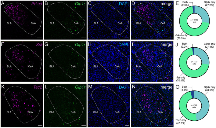
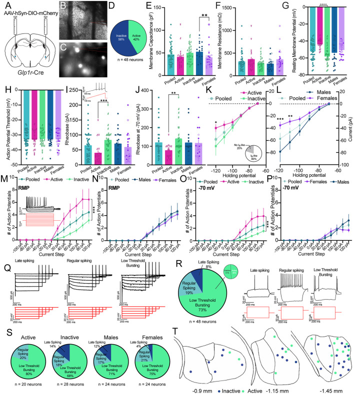
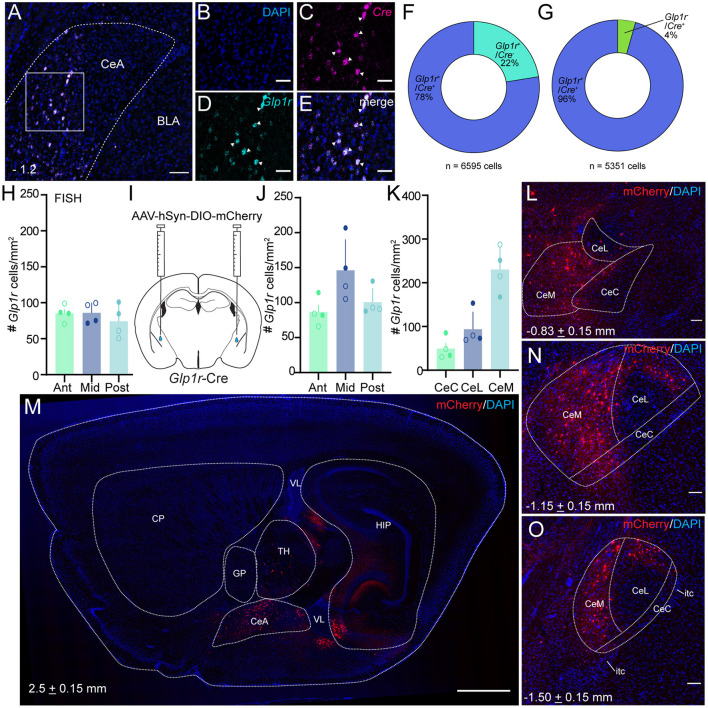
Glucagon-like peptide 1 receptors (GLP-1Rs) are highly expressed in the brain and are responsible for mediating the acute anorexigenic actions of widely prescribed GLP-1R agonists. Neurobiological efforts to localize the hypophagic effects of GLP-1R agonists in the brain have mainly focused on the hypothalamus and hindbrain. In this study, we performed a deep anatomical and neurophysiological characterization of GLP-1Rs in the central nucleus of the amygdala (CeA). At an mRNA level, we found that is diffusely coexpressed in known CeA subpopulations like protein kinase c δ (), somatostatin (), or tachykinin2 (). At a cellular level, we used Cre mice and viral Cre-dependent tracing to map the anatomical positions of GLP-1R cells across the rostral-caudal axis of the CeA and in CeA subdivisions. We found that 1 cells are highly enriched in the medial subdivision of the CeA (CeM). Using whole cell patch clamp electrophysiology, we found that 1 neurons are characterized by the presence of I-like currents and resemble a low threshold bursting neuronal subtype in response to hyperpolarizing and depolarizing current injections. We observed sex differences in the magnitude of I-like currents and membrane capacitance. At rest, we observed that nearly half of 1 neurons are spontaneously active. We observed that active and inactive neurons display significant differences in excitability even when normalized to an identical holding potential. Our data are the first to deeply characterize the pattern of in the CeA and study the neurophysiological characteristics of CeA neurons expressing . Future studies leveraging these data will be important to understanding the impact of GLP-1R agonists on feeding and motivation.
胰高血糖素样肽1受体(GLP-1Rs)在大脑中高度表达,负责介导广泛使用的GLP-1R激动剂的急性厌食作用。将GLP-1R激动剂的促食欲作用定位到大脑中的神经生物学研究主要集中在下丘脑和后脑。在本研究中,我们对杏仁核中央核(CeA)中的GLP-1Rs进行了深入的解剖学和神经生理学特征分析。在mRNA水平上,我们发现其在已知的CeA亚群如蛋白激酶cδ()、生长抑素()或速激肽2()中广泛共表达。在细胞水平上,我们使用Cre小鼠和病毒Cre依赖性追踪来绘制CeA整个头尾轴和CeA细分区域中GLP-1R细胞的解剖位置。我们发现CeA内侧细分区域(CeM)中GLP-1R细胞高度富集。使用全细胞膜片钳电生理学,我们发现GLP-1R神经元的特征是存在I类电流,并且在响应超极化和去极化电流注入时类似于低阈值爆发神经元亚型。我们观察到I类电流幅度和膜电容存在性别差异。在静息状态下,我们观察到近一半的GLP-1R神经元自发活动。我们观察到,即使将活性和非活性神经元归一化到相同的钳制电位,它们在兴奋性上也存在显著差异。我们的数据首次深入表征了CeA中GLP-1R的模式,并研究了表达GLP-1R的CeA神经元的神经生理学特征。利用这些数据的未来研究对于理解GLP-1R激动剂对进食和动机的影响将非常重要。