Nakatani Eri, Okajima Riho, Ohnuma Kiyoshi
Department of Science of Technology Innovation, Nagaoka University of Technology, 1603-1, Kamitomioka-machi, Nagaoka, 940-2188, Japan.
Department of Bioengineering, Nagaoka University of Technology, 1603-1, Kamitomioka-machi, Nagaoka, 940-2188, Japan.
Biochem Biophys Rep. 2021 Dec 27;29:101195. doi: 10.1016/j.bbrep.2021.101195. eCollection 2022 Mar.
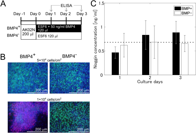
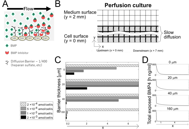
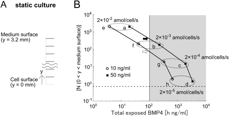
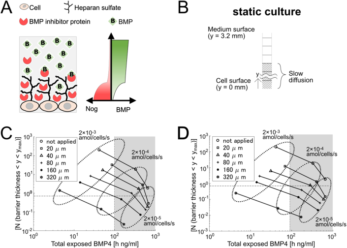
Auto/paracrine factors secreted from cells affect differentiation of human pluripotent stem cells (hPSCs). However, the molecular mechanisms underlying the role of secreted factors are not well known. We previously showed that pattern formation in hPSCs induced by BMP4 could be reproduced by a simple reaction-diffusion of BMP and Noggin, a cell-secreted BMP4 inhibitor. However, the amount of Noggin secreted is unknown. In this study, we measured the concentration of Noggin secreted during the differentiation of hPSCs induced by BMP4. The Noggin concentration in the supernatant before and after differentiation was constant at approximately 0.69 ng/mL, which is approximately 50-200 times less than expected in the model. To explain the difference between the experiment and model, we assumed that macromolecules such as heparan sulfate proteoglycan on the cell surface act as a diffusion barrier structure, where the diffusion slows down to 1/400. The model with the diffusion barrier structure reduced the Noggin concentration required to suppress differentiation in the static culture model. The model also qualitatively reproduced the pattern formation, in which only the upstream but not the downstream hPSCs were differentiated in a one-directional perfusion culture chamber, with a small change in the amount of secreted Noggin resulting in a large change in the differentiation position. These results suggest that the diffusion barrier on the cell surface might enhance the auto/paracrine effects on monolayer hPSC culture.
细胞分泌的自分泌/旁分泌因子会影响人类多能干细胞(hPSC)的分化。然而,分泌因子发挥作用的分子机制尚不清楚。我们之前表明,BMP4诱导的hPSC中的模式形成可以通过BMP和Noggin(一种细胞分泌的BMP4抑制剂)的简单反应扩散来重现。然而,Noggin的分泌量尚不清楚。在本研究中,我们测量了BMP4诱导hPSC分化过程中分泌的Noggin的浓度。分化前后上清液中的Noggin浓度恒定在约0.69 ng/mL,这比模型中预期的浓度低约50 - 200倍。为了解释实验与模型之间的差异,我们假设细胞表面的大分子如硫酸乙酰肝素蛋白聚糖作为扩散屏障结构,扩散速度减慢至1/400。具有扩散屏障结构的模型降低了静态培养模型中抑制分化所需的Noggin浓度。该模型还定性地重现了模式形成,即在单向灌注培养室中,只有上游而非下游的hPSC发生分化,分泌的Noggin量的微小变化会导致分化位置的巨大变化。这些结果表明,细胞表面的扩散屏障可能会增强对单层hPSC培养的自分泌/旁分泌作用。