Rush Alzheimer's Disease Center, Rush University Medical Center, 1750 W Harrison Street, Chicago, IL, 60612, USA.
Department of Pathology, Rush University Medical Center, Chicago, IL, USA.
Acta Neuropathol. 2022 Mar;143(3):349-362. doi: 10.1007/s00401-021-02397-x. Epub 2022 Jan 19.
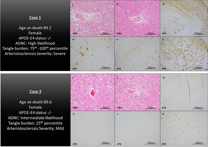
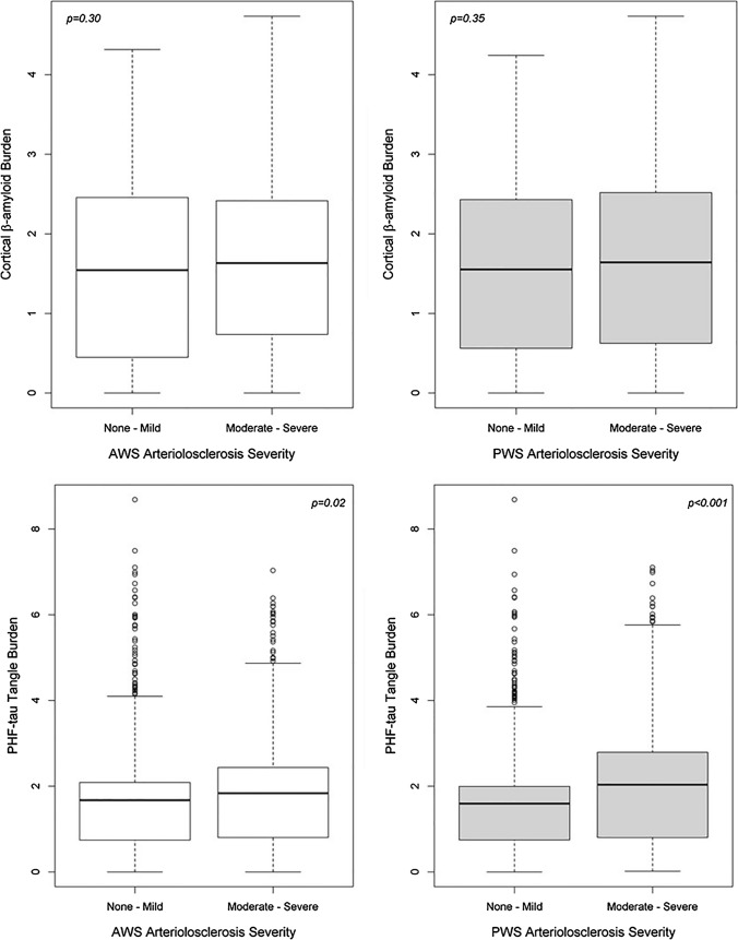
Emerging evidence suggests that small vessel disease (SVD) is a risk factor for clinical dementia and may contribute to AD neuropathological changes. Watershed brain regions are located at the most distal areas between arterial territories, making them vulnerable to SVD-related changes. We examined the association of pathologic markers of SVD, specifically arteriolosclerosis in watershed brain regions, with AD pathologic changes. Participants (N = 982; mean age-at-death = 90; 69% women) were enrolled as part of one of two cohort studies of aging and dementia. At autopsy, neuropathological evaluation included semi-quantitative grading of arteriolosclerosis pathology from 2 cortical watershed regions: the anterior watershed (AWS) and posterior watershed (PWS), densities for cortical β-amyloid and tau-tangle pathology, and other common age-related pathologies. Linear regression models examined the association of watershed arteriolosclerosis pathology with β-amyloid and tau-tangle burden. In follow-up analyses, available ex-vivo MRI and proteomics data in a subset of decedents were leveraged to examine the association of whole brain measure of WMH, as a presumed MRI marker of SVD, with β-amyloid and tau-tangle burden, as well as to examine the association of watershed arteriolosclerosis with proteomic tau. Watershed arteriolosclerosis was common, with 45% of older persons having moderate-to-severe arteriolosclerosis pathology in the AWS region, and 35% in the PWS. In fully adjusted models that controlled for demographics and common age-related pathologies, an increase in severity of PWS arteriolosclerosis was associated with a higher burden of tau-tangle burden, specifically neocortical tau burden, but not with β-amyloid. AWS arteriolosclerosis was not associated with β-amyloid or tau pathology. Ex-vivo WMH was associated with greater tau-tangle pathology burden but not β-amyloid. Furthermore, PWS arteriolosclerosis was associated with higher abundance of tau phosphopeptides, that promote formation of tau aggregates. These data provide compelling evidence that SVD, specifically posterior watershed arteriolosclerosis pathology, is linked with tau pathological changes in the aging brain.
新出现的证据表明,小血管疾病 (SVD) 是临床痴呆的一个危险因素,并且可能导致 AD 神经病理学变化。分水岭脑区位于动脉供血区之间最远端的部位,因此容易受到 SVD 相关变化的影响。我们研究了 SVD 的病理标志物,特别是分水岭脑区的小动脉硬化,与 AD 病理变化的关系。参与者 (N=982; 死亡时的平均年龄=90; 69%为女性) 作为老龄化和痴呆症的两项队列研究之一的一部分被招募。在尸检时,神经病理学评估包括从 2 个皮质分水岭区 (AWS 和 PWS) 对小动脉硬化病理学进行半定量分级、皮质β-淀粉样蛋白和 tau 缠结病理学的密度,以及其他常见的与年龄相关的病理学。线性回归模型检查了分水岭小动脉硬化病理学与β-淀粉样蛋白和 tau 缠结负担的关系。在后续分析中,利用部分死者的可获得的离体 MRI 和蛋白质组学数据,研究了全脑白质高信号 (WMH) 的测量值,作为 SVD 的假定 MRI 标志物,与β-淀粉样蛋白和 tau 缠结负担的关系,以及研究分水岭小动脉硬化与蛋白质组学 tau 的关系。分水岭小动脉硬化很常见,45%的老年人 AWS 区域存在中度至重度小动脉硬化病理学,35%的老年人 PWS 区域存在中度至重度小动脉硬化病理学。在完全调整了人口统计学和常见与年龄相关的病理学的模型中,PWS 小动脉硬化严重程度的增加与 tau 缠结负担的增加相关,特别是新皮质 tau 负担的增加,但与β-淀粉样蛋白无关。AWS 小动脉硬化与β-淀粉样蛋白或 tau 病理学无关。离体 WMH 与 tau 缠结病理学负担增加相关,但与β-淀粉样蛋白无关。此外,PWS 小动脉硬化与促进 tau 聚集的 tau 磷酸肽的丰度增加有关。这些数据提供了令人信服的证据,表明 SVD,特别是后分水岭小动脉硬化病理学,与衰老大脑中的 tau 病理变化有关。