Institute for Fundamental Biomedical Research, Johns Hopkins All Children's Hospital, St. Petersburg, Florida, USA.
Division of Endocrinology, Diabetes and Metabolism, Department of Medicine, Johns Hopkins University School of Medicine, Baltimore, Maryland, USA.
JCI Insight. 2022 Mar 22;7(6):e153136. doi: 10.1172/jci.insight.153136.
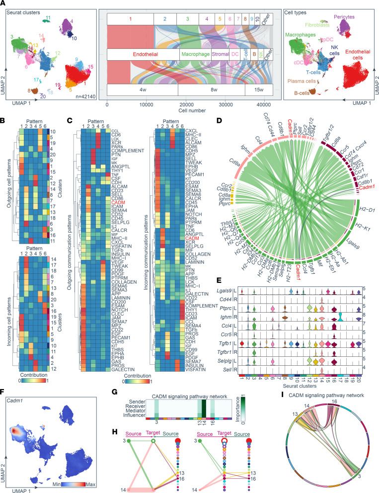
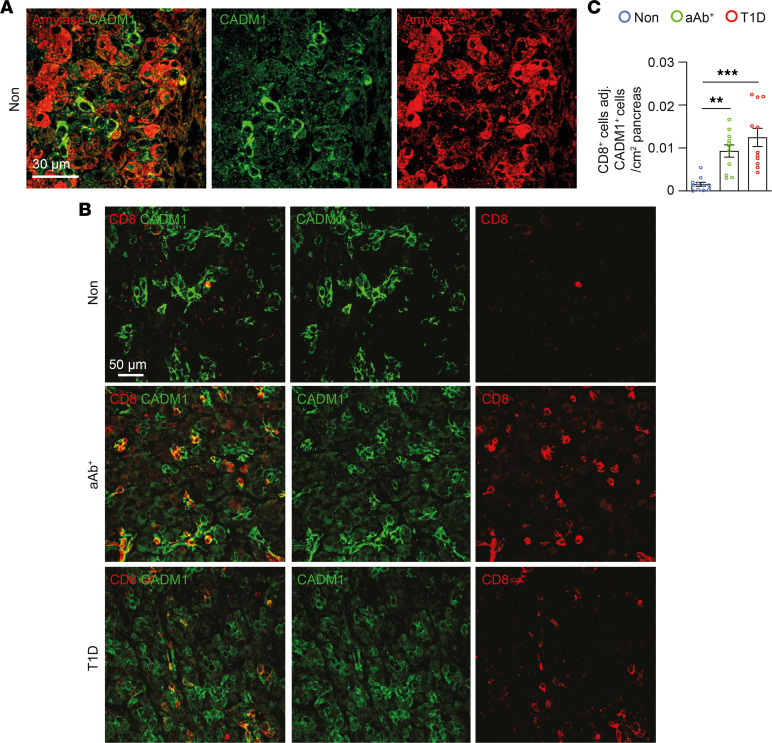
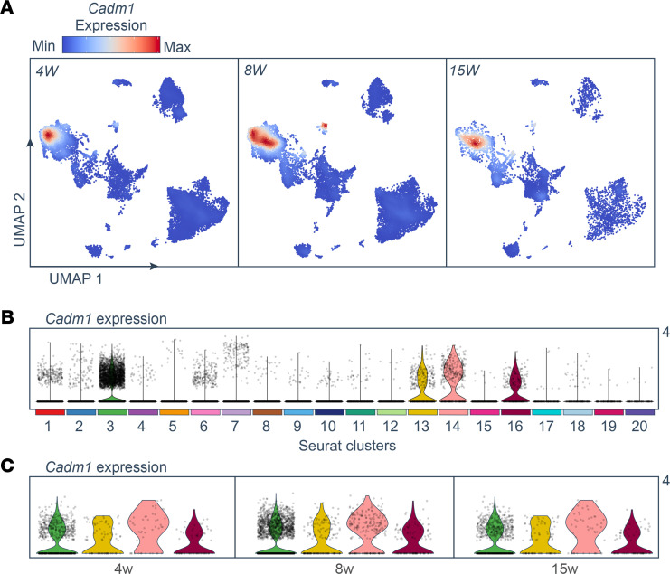
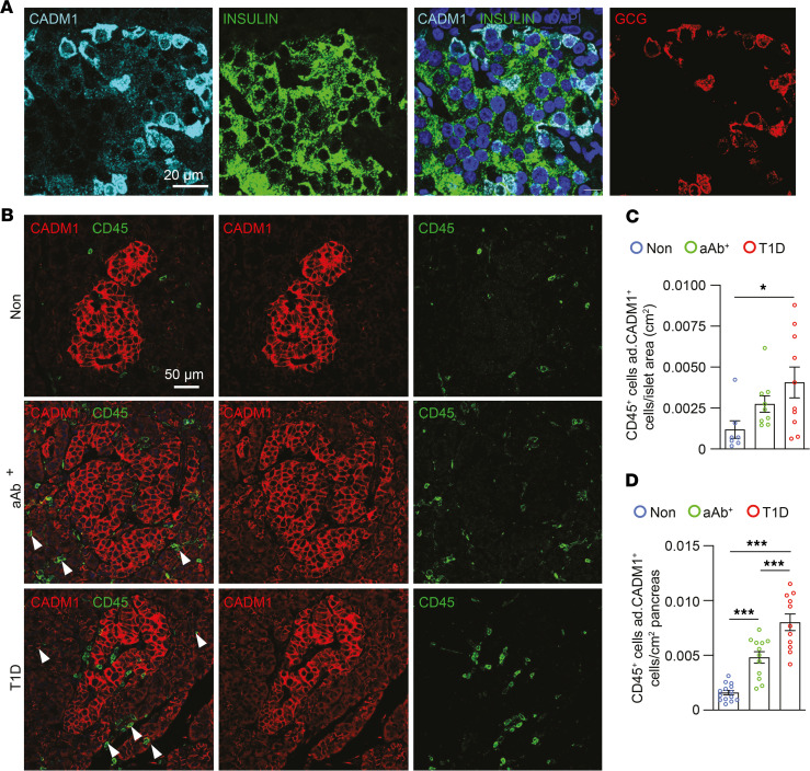
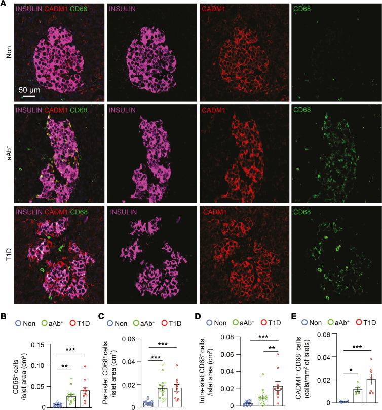
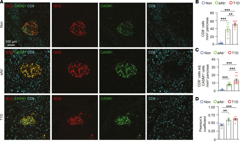
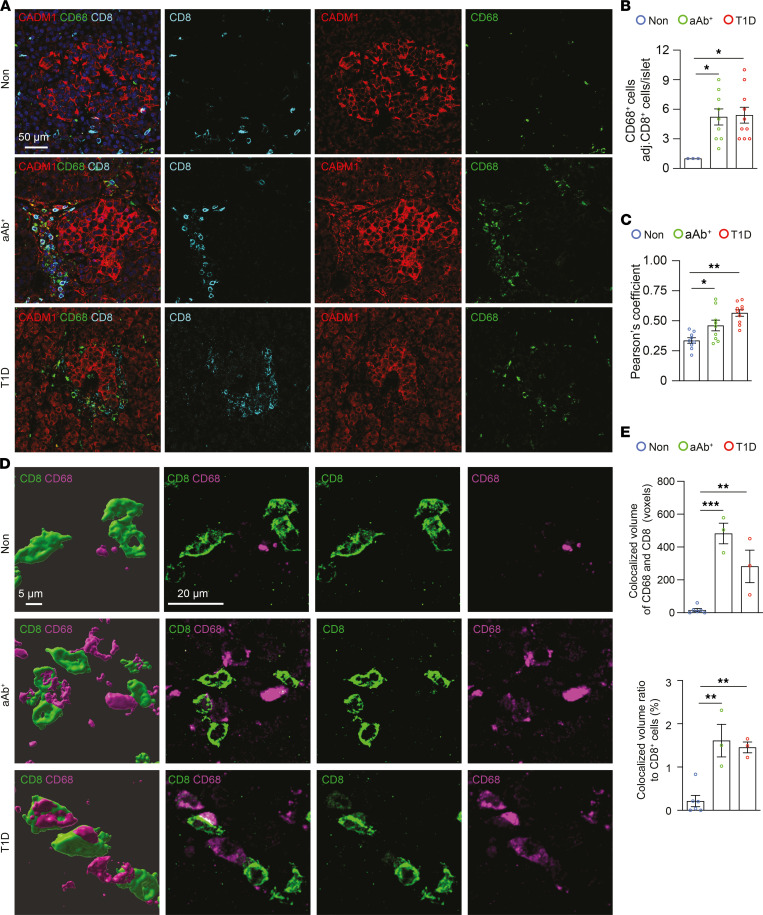
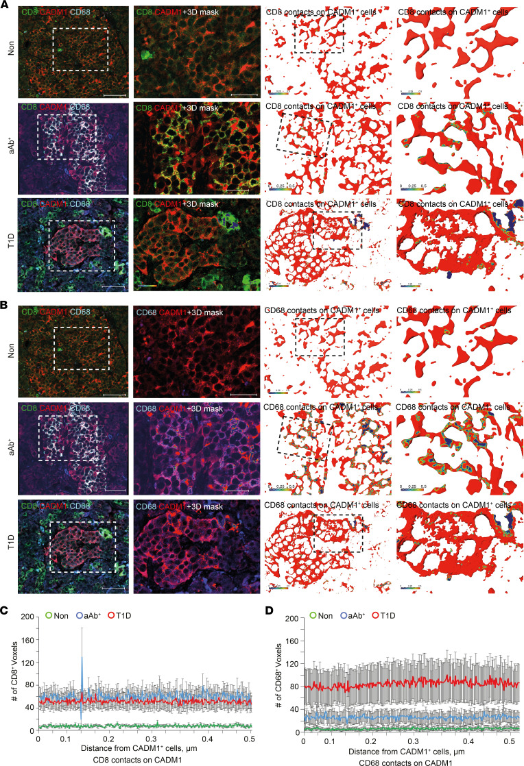
BACKGROUNDPathophysiology of type 1 diabetes (T1D) is illustrated by pancreatic islet infiltration of inflammatory lymphocytes, including CD8+ T cells; however, the molecular factors mediating their recruitment remain unknown. We hypothesized that single-cell RNA-sequencing (scRNA-Seq) analysis of immune cell populations isolated from islets of NOD mice captured gene expression dynamics providing critical insight into autoimmune diabetes pathogenesis.METHODSPancreatic sections from human donors were investigated, including individuals with T1D, autoantibody-positive (aAb+) individuals, and individuals without diabetes who served as controls. IHC was performed to assess islet hormones and both novel and canonical immune cell markers that were identified from unbiased, state-of-the-art workflows after reanalyzing murine scRNA-Seq data sets.RESULTSComputational workflows identified cell adhesion molecule 1-mediated (Cadm1-mediated) homotypic binding among the most important intercellular interactions among all cell clusters, as well as Cadm1 enrichment in macrophages and DCs from pancreata of NOD mice. Immunostaining of human pancreata revealed an increased number of CADM1+glucagon+ cells adjacent to CD8+ T cells in sections from T1D and aAb+ donors compared with individuals without diabetes. Numbers of CADM1+CD68+ peri-islet myeloid cells adjacent to CD8+ T cells were also increased in pancreatic sections from both T1D and aAb+ donors compared with individuals without diabetes.CONCLUSIONIncreased detection of CADM1+ cells adjacent to CD8+ T cells in pancreatic sections of individuals with T1D and those who were aAb+ validated workflows and indicated CADM1-mediated intercellular contact may facilitate islet infiltration of cytotoxic T lymphocytes and serve as a potential therapeutic target for preventing T1D pathogenesis.FUNDINGThe Johns Hopkins All Children's Foundation Institutional Research Grant Program, the National Natural Science Foundation of China (grant 82071326), and the Deutsche Forschungsgemeinschaft (grants 431549029-SFB1451, EXC2030-390661388, and 411422114-GRK2550).
1 型糖尿病(T1D)的病理生理学表现为胰岛浸润炎症性淋巴细胞,包括 CD8+T 细胞;然而,介导其募集的分子因素仍不清楚。我们假设,对从 NOD 小鼠胰岛中分离的免疫细胞群体进行单细胞 RNA 测序(scRNA-Seq)分析,可以捕获提供关键见解的基因表达动态自身免疫性糖尿病发病机制。
研究了来自人类供体的胰腺切片,包括 T1D 患者、自身抗体阳性(aAb+)个体和无糖尿病对照个体。进行免疫组织化学染色以评估胰岛激素和新型和经典免疫细胞标志物,这些标志物是从重新分析鼠 scRNA-Seq 数据集后使用无偏、最先进的工作流程识别的。
计算工作流程确定了细胞间相互作用中最重要的细胞间相互作用之一,即细胞间粘附分子 1 介导的(Cadm1 介导的)同种型结合,以及 NOD 小鼠胰腺中的巨噬细胞和 DC 中的 Cadm1 富集。免疫染色显示,与无糖尿病个体相比,T1D 和 aAb+供体胰腺切片中 CD8+T 细胞附近的 CADM1+胰高血糖素+细胞数量增加。与无糖尿病个体相比,T1D 和 aAb+供体胰腺切片中 CD8+T 细胞附近的 CADM1+CD68+胰岛周围髓样细胞数量也增加。
在 T1D 个体和 aAb+个体的胰腺切片中,与 CD8+T 细胞相邻的 CADM1+细胞的检测增加验证了工作流程,并表明 CADM1 介导的细胞间接触可能有助于细胞毒性 T 淋巴细胞浸润胰岛,并可作为预防 T1D 发病机制的潜在治疗靶点。
约翰霍普金斯所有儿童基金会机构研究赠款计划、中国国家自然科学基金(资助 82071326)和德国研究基金会(资助 431549029-SFB1451、EXC2030-390661388 和 411422114-GRK2550)。