• 文献检索
  • 文档翻译
  • 深度研究
  • 学术资讯
  • Suppr Zotero 插件Zotero 插件
  • 邀请有礼
  • 套餐&价格
  • 历史记录
应用&插件
Suppr Zotero 插件Zotero 插件浏览器插件Mac 客户端Windows 客户端微信小程序
定价
高级版会员购买积分包购买API积分包
服务
文献检索文档翻译深度研究API 文档MCP 服务
关于我们
关于 Suppr公司介绍联系我们用户协议隐私条款
关注我们

Suppr 超能文献

核心技术专利:CN118964589B侵权必究
粤ICP备2023148730 号-1Suppr @ 2026

文献检索

告别复杂PubMed语法,用中文像聊天一样搜索,搜遍4000万医学文献。AI智能推荐,让科研检索更轻松。

立即免费搜索

文件翻译

保留排版,准确专业,支持PDF/Word/PPT等文件格式,支持 12+语言互译。

免费翻译文档

深度研究

AI帮你快速写综述,25分钟生成高质量综述,智能提取关键信息,辅助科研写作。

立即免费体验

CT扫描对肺裂解剖变异的研究

Study on Anatomical Variations in Fissures of Lung by CT Scan.

作者信息

Manjunath M, Sharma M Vishnu, Janso Kollanur, John Praveen Kumar, Anupama N, Harsha D S

机构信息

Department of Respiratory Medicine, Navodaya Medical College, Raichur, Karnataka, India.

Department of Respiratory Medicine, A.J. Institute of Medical Sciences and Research Center, Kuntikana, Mangalore, Karnataka, India.

出版信息

Indian J Radiol Imaging. 2022 Jan 11;31(4):797-804. doi: 10.1055/s-0041-1741045. eCollection 2021 Oct.

DOI:10.1055/s-0041-1741045
PMID:35136490
原文链接:https://pmc.ncbi.nlm.nih.gov/articles/PMC8817794/
Abstract

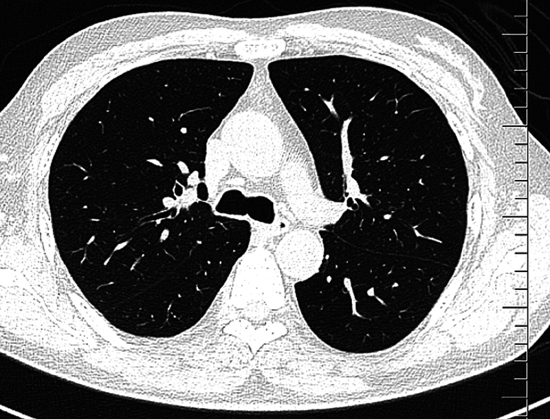
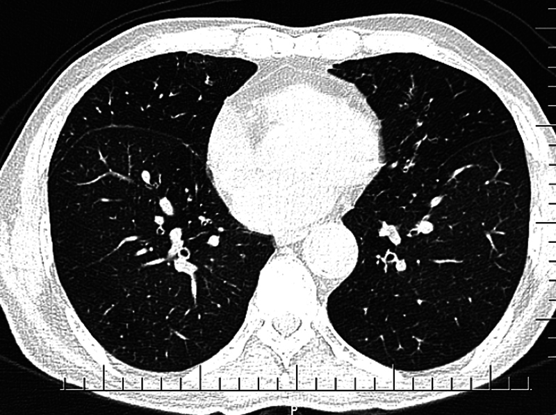
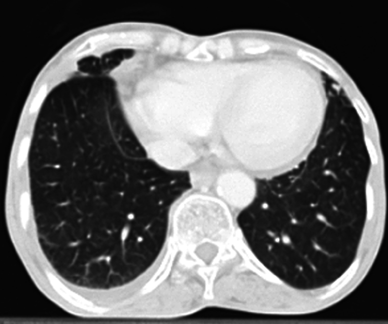
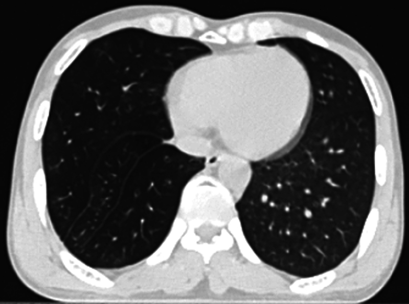
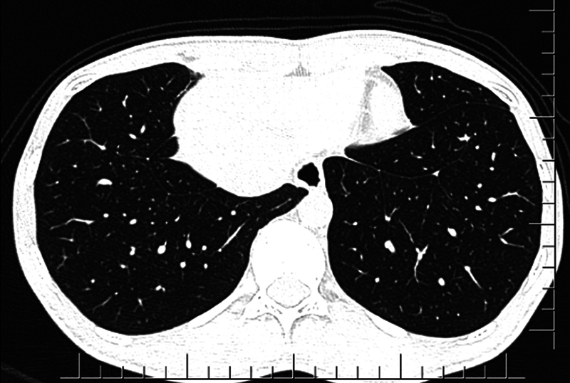
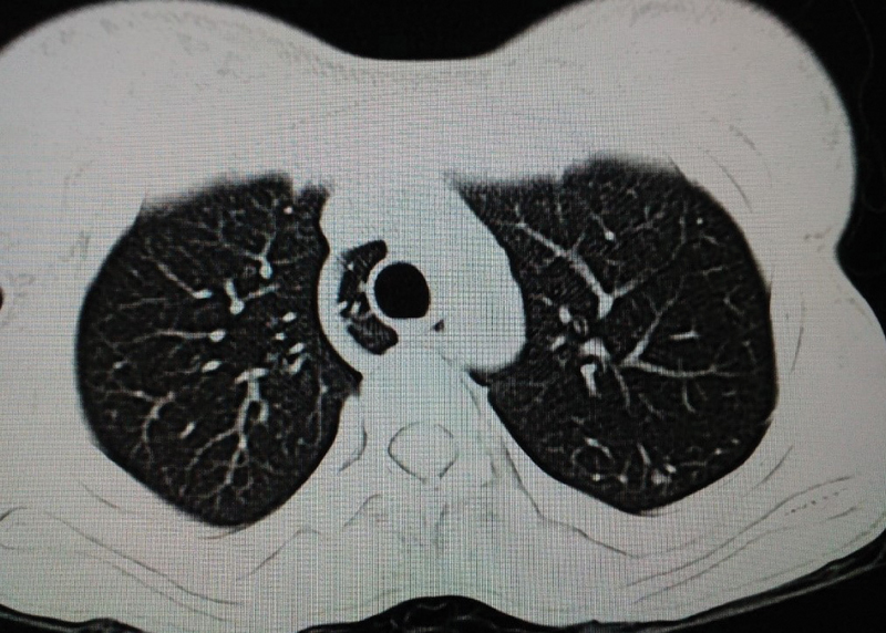
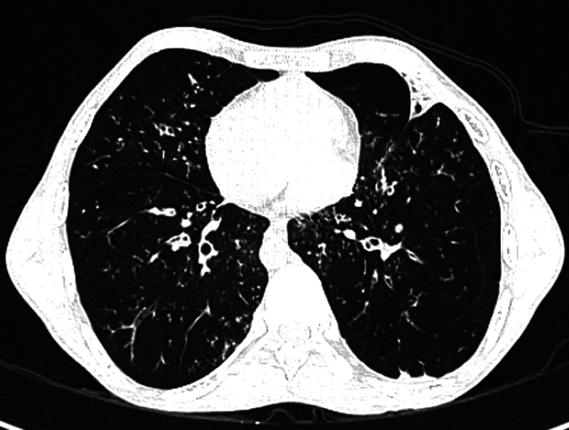
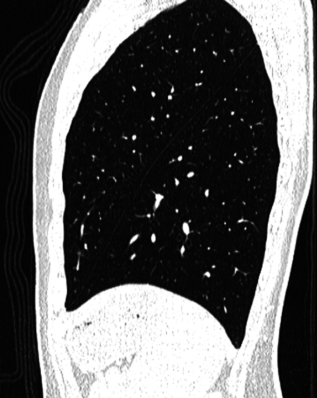
Refinements in the modern computed tomography (CT) imaging techniques have led to anatomical variations in the fissures of lung being diagnosed more frequently. So far, majority of the studies conducted are cadaveric. There is paucity of studies in this aspect based on chest CT images. Hence, we undertook this study to find the anatomical variations in the fissures. Prior detection of anatomical abnormalities is important to reduce postoperative complications in lung resection surgeries.  This was a cross-sectional study conducted over a period of 2 years. Data were collected from the patients who underwent CT scan thorax. Patients in whom normal anatomy of lung was distorted and cases where both lungs were not visualized completely were excluded from the study. All the CT images were reviewed by a single radiologist. The presence or absence of the normal and accessory pulmonary fissures, as well as the continuity of each fissure, was recorded by the radiologist. Data were compiled and analyzed.  The study population consisted of 394 (70.4%) males and 166 (29.6%) females, totaling 560 cases. Fissural variations were detected in 22.9% (  = 128). Also, 17.5% (  = 98) fissural variations were seen in males and 5.4% (  = 30) fissural variations were seen in females. Further, 54.7% (  = 70) of variations were detected in the right lung and 45.3% (  = 58) in the left lung. The most common fissural variation noted was right incomplete oblique fissure with a frequency of 8.4% cases (  = 47). The most common accessory fissure detected was inferior accessory fissure. Total 22 cases were detected in both the lungs, 17 cases in male and 5 in female.  Anatomical variations in fissures were found to be more in the right lung than the left lung. Accessory fissures were detected in higher incidence on the right side.

摘要

现代计算机断层扫描(CT)成像技术的改进使得肺部裂的解剖变异被更频繁地诊断出来。到目前为止,大多数相关研究是基于尸体解剖的。基于胸部CT图像在这方面的研究较少。因此,我们开展这项研究以发现肺部裂的解剖变异。术前检测解剖异常对于减少肺切除手术的术后并发症很重要。

这是一项为期2年的横断面研究。数据收集自接受胸部CT扫描的患者。肺部解剖结构正常但被扭曲以及双肺未完全显影的患者被排除在研究之外。所有CT图像均由一名放射科医生进行评估。放射科医生记录正常和副肺裂的存在与否以及每个裂的连续性。数据进行整理和分析。

研究人群包括394名男性(70.4%)和166名女性(29.6%),共560例。发现22.9%(n = 128)存在裂变异。其中,男性中裂变异的发生率为17.5%(n = 98),女性为5.4%(n = 30)。此外,右肺变异占54.7%(n = 70),左肺占45.3%(n = 58)。最常见的裂变异是右斜裂不完全,发生率为8.4%(n = 47)。最常见的副裂是下副裂。双肺共检测到22例,其中男性17例,女性5例。

发现肺部裂的解剖变异在右肺比左肺更多见。副裂在右侧的发生率更高。

https://cdn.ncbi.nlm.nih.gov/pmc/blobs/be06/8817794/9ab0423b0a4e/10-1055-s-0041-1741045-i56120-10.jpg
https://cdn.ncbi.nlm.nih.gov/pmc/blobs/be06/8817794/44f3bcc0c307/10-1055-s-0041-1741045-i56120-1.jpg
https://cdn.ncbi.nlm.nih.gov/pmc/blobs/be06/8817794/0ecb1b5b12c2/10-1055-s-0041-1741045-i56120-2.jpg
https://cdn.ncbi.nlm.nih.gov/pmc/blobs/be06/8817794/8c12860efb7d/10-1055-s-0041-1741045-i56120-3.jpg
https://cdn.ncbi.nlm.nih.gov/pmc/blobs/be06/8817794/22bace857dd9/10-1055-s-0041-1741045-i56120-4.jpg
https://cdn.ncbi.nlm.nih.gov/pmc/blobs/be06/8817794/3ef6af5d7b24/10-1055-s-0041-1741045-i56120-5.jpg
https://cdn.ncbi.nlm.nih.gov/pmc/blobs/be06/8817794/cd99db6bf014/10-1055-s-0041-1741045-i56120-6.jpg
https://cdn.ncbi.nlm.nih.gov/pmc/blobs/be06/8817794/82898f67d867/10-1055-s-0041-1741045-i56120-7.jpg
https://cdn.ncbi.nlm.nih.gov/pmc/blobs/be06/8817794/c4ae0a2c1f14/10-1055-s-0041-1741045-i56120-8.jpg
https://cdn.ncbi.nlm.nih.gov/pmc/blobs/be06/8817794/a533106507d0/10-1055-s-0041-1741045-i56120-9.jpg
https://cdn.ncbi.nlm.nih.gov/pmc/blobs/be06/8817794/9ab0423b0a4e/10-1055-s-0041-1741045-i56120-10.jpg

相似文献

1
Study on Anatomical Variations in Fissures of Lung by CT Scan.CT扫描对肺裂解剖变异的研究
Indian J Radiol Imaging. 2022 Jan 11;31(4):797-804. doi: 10.1055/s-0041-1741045. eCollection 2021 Oct.
2
Anatomical Variations in Pulmonary Fissures on Computed Tomography (CT).计算机断层扫描(CT)上肺裂的解剖变异
Cureus. 2022 Nov 30;14(11):e32062. doi: 10.7759/cureus.32062. eCollection 2022 Nov.
3
Variations in pulmonary fissural anatomy: a medicolegal autopsy study of 256 cases.肺裂解剖变异:256例法医尸检研究
ANZ J Surg. 2020 Apr;90(4):608-611. doi: 10.1111/ans.15553. Epub 2019 Nov 10.
4
Are textbook lungs really normal? A cadaveric study on the anatomical and clinical importance of variations in the major lung fissures, and the incomplete right horizontal fissure.教科书肺真的正常吗?一项关于主要肺裂和不完全右水平裂在解剖学和临床重要性的大体研究。
Clin Anat. 2021 Apr;34(3):387-396. doi: 10.1002/ca.23661. Epub 2020 Aug 17.
5
Preoperative delineation of pulmonary fissural anatomy at multi-detector computed tomography in children with congenital pulmonary malformations and impact on surgical complications and postoperative course.多排螺旋 CT 术前对先天性肺畸形患儿肺裂解剖结构的描绘及其对手术并发症和术后病程的影响。
Pediatr Radiol. 2020 May;50(5):636-645. doi: 10.1007/s00247-020-04618-z. Epub 2020 Jan 28.
6
Variations in human pulmonary fissures and lobes: a study conducted in nepalese cadavers.人类肺裂和肺叶的变异:在尼泊尔尸体上进行的一项研究。
Anat Cell Biol. 2018 Jun;51(2):85-92. doi: 10.5115/acb.2018.51.2.85. Epub 2018 Jun 27.
7
MAJOR ANATOMIC VARIATIONS OF PULMONARY FISSURES AND LOBES ON POSTMORTEM EXAMINATION.尸体解剖时肺裂和肺叶的主要解剖变异
Acta Clin Croat. 2015 Jun;54(2):201-7.
8
Pulmonary fissures and lobes: a cadaveric study with emphasis on surgical and radiological implications.肺裂和肺叶:一项侧重于手术及放射学意义的尸体研究
Clin Ter. 2012;163(1):9-13.
9
Incomplete and accessory fissures of the lung evaluated by high-resolution computed tomography.高分辨率计算机断层扫描评估肺的不完整和附属裂。
Eur J Radiol. 2014 Mar;83(3):595-9. doi: 10.1016/j.ejrad.2013.12.001. Epub 2013 Dec 12.
10
Accessory fissures of the upper lobe of the left lung: CT and plain film appearance.左肺上叶副裂:CT及平片表现
AJR Am J Roentgenol. 1994 Jun;162(6):1287-93. doi: 10.2214/ajr.162.6.8191982.

引用本文的文献

1
Lung lobe segmentation: performance of open-source MOOSE, TotalSegmentator, and LungMask models compared to a local in-house model.肺叶分割:与本地内部模型相比,开源MOOSE、TotalSegmentator和LungMask模型的性能
Eur Radiol Exp. 2025 Sep 4;9(1):86. doi: 10.1186/s41747-025-00623-9.
2
Morphological variations of fissures, lobes, and hilar pattern of the lung in a select South African sample.选择南非样本中的肺裂、叶和肺门模式的形态学变异。
Surg Radiol Anat. 2024 Dec;46(12):2005-2017. doi: 10.1007/s00276-024-03497-5. Epub 2024 Oct 10.
3
A pulmonary nodule mislocated in "dorsal" segment due to tri-lobed left lung.

本文引用的文献

1
Morphological variations of the lungs: a study conducted on Indian cadavers.肺的形态学变异:对印度尸体进行的一项研究。
Anat Cell Biol. 2014 Dec;47(4):253-8. doi: 10.5115/acb.2014.47.4.253. Epub 2014 Dec 23.
2
Incomplete and accessory fissures of the lung evaluated by high-resolution computed tomography.高分辨率计算机断层扫描评估肺的不完整和附属裂。
Eur J Radiol. 2014 Mar;83(3):595-9. doi: 10.1016/j.ejrad.2013.12.001. Epub 2013 Dec 12.
3
The National Emphysema Treatment Trial (NETT) Part II: Lessons learned about lung volume reduction surgery.
由于左肺呈三叶状,一个肺结节位于“背侧”段。
Front Surg. 2023 Jan 6;9:1069543. doi: 10.3389/fsurg.2022.1069543. eCollection 2022.
4
Anatomical Variations in Pulmonary Fissures on Computed Tomography (CT).计算机断层扫描(CT)上肺裂的解剖变异
Cureus. 2022 Nov 30;14(11):e32062. doi: 10.7759/cureus.32062. eCollection 2022 Nov.
国家肺气肿治疗试验(NETT)第二部分:肺减容手术的经验教训。
Am J Respir Crit Care Med. 2011 Oct 15;184(8):881-93. doi: 10.1164/rccm.201103-0455CI.
4
Lung morphology: a cadaver study in Indian population.肺形态学:一项针对印度人群的尸体研究。
Ital J Anat Embryol. 2010;115(3):235-40.
5
Migrating azygos vein and vanishing azygos lobe: MDCT findings.奇静脉移位和奇静脉叶消失:MDCT 表现。
AJR Am J Roentgenol. 2010 Mar;194(3):599-603. doi: 10.2214/AJR.09.3303.
6
Normal and accessory fissures of the lung: evaluation with contiguous volumetric thin-section multidetector CT.肺的正常和副裂:连续容积薄层多排 CT 评估。
Eur J Radiol. 2010 Aug;75(2):e1-8. doi: 10.1016/j.ejrad.2009.10.009. Epub 2009 Nov 7.
7
Evaluation of pulmonary lobe variations using multidetector row computed tomography.使用多排探测器计算机断层扫描评估肺叶变异
J Comput Assist Tomogr. 2007 Nov-Dec;31(6):956-60. doi: 10.1097/rct.0b013e3180500d23.
8
The approach of fused fissures with fissureless technique decreases the incidence of persistent air leak after lobectomy.采用无裂隙技术融合裂隙可降低肺叶切除术后持续性漏气的发生率。
Eur J Cardiothorac Surg. 2007 Feb;31(2):203-8. doi: 10.1016/j.ejcts.2006.11.030. Epub 2006 Dec 18.
9
Variability of the pulmonary oblique fissures presented by high-resolution computed tomography.高分辨率计算机断层扫描显示的肺斜裂变异
Surg Radiol Anat. 2006 Jun;28(3):293-9. doi: 10.1007/s00276-006-0079-y. Epub 2006 Feb 7.
10
Morphological variations of the lung fissures and lobes.肺裂和肺叶的形态学变异。
Indian J Chest Dis Allied Sci. 2004 Jul-Sep;46(3):179-82.