Niehues Hanna, Rikken Gijs, van Vlijmen-Willems Ivonne M J J, Rodijk-Olthuis Diana, van Erp Piet E J, Zeeuwen Patrick L J M, Schalkwijk Joost, van den Bogaard Ellen H
Department of Dermatology, Radboud University Medical Center (Radboudumc), Radboud Institute for Molecular Life Sciences (RIMLS), Nijmegen, The Netherlands.
JID Innov. 2021 Oct 22;2(1):100066. doi: 10.1016/j.xjidi.2021.100066. eCollection 2022 Jan.
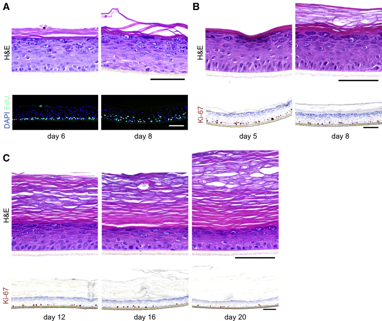
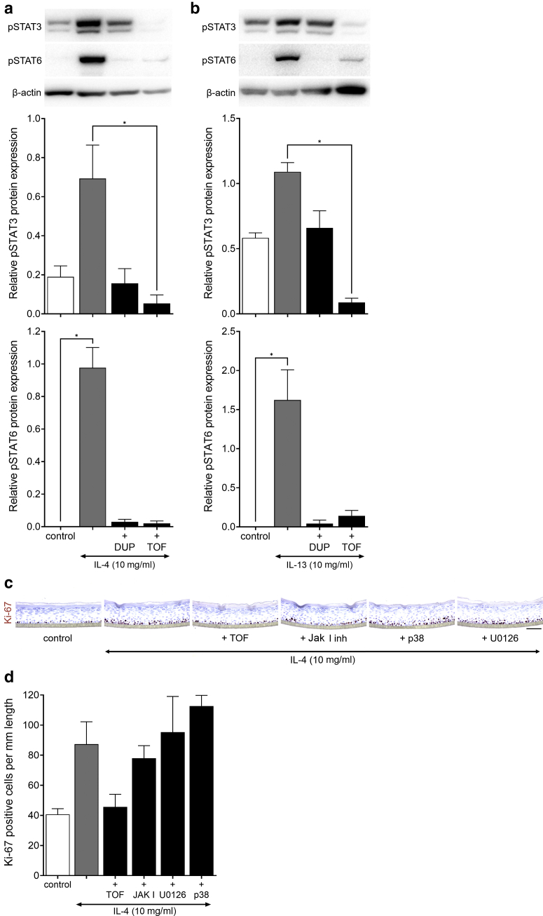
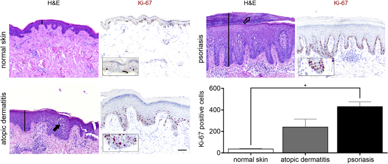
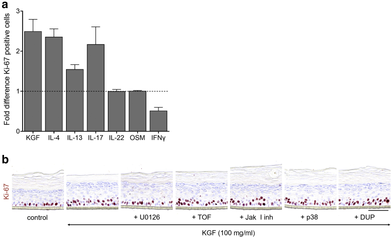
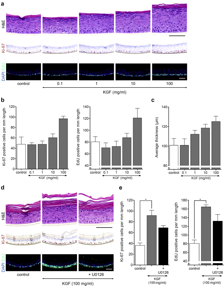
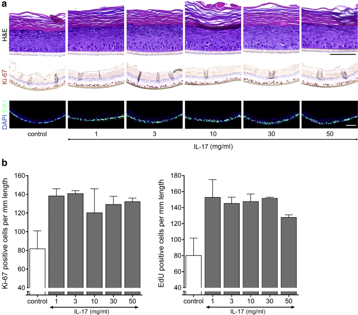
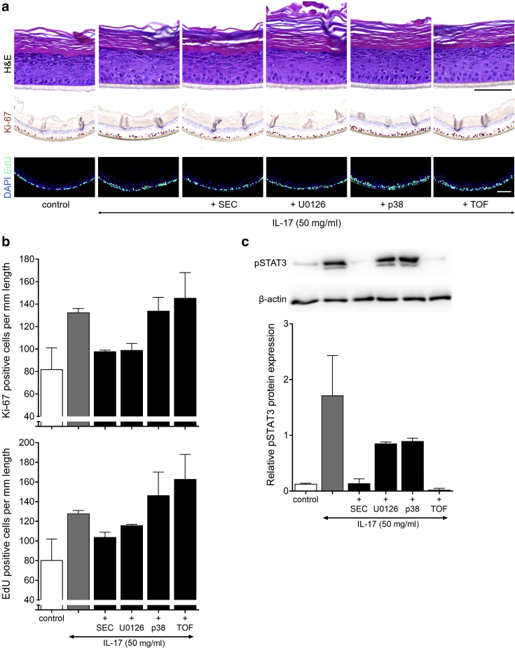
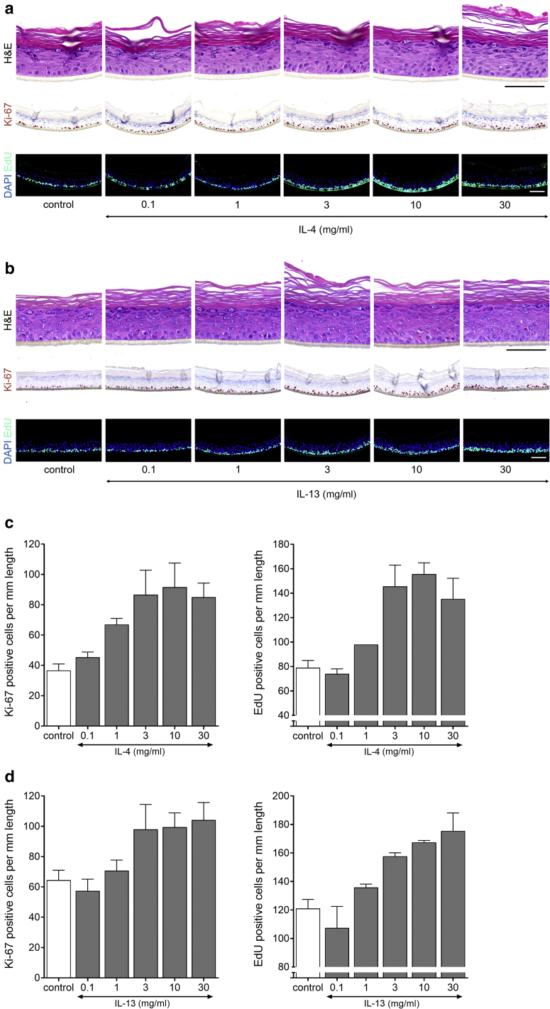
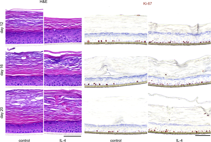
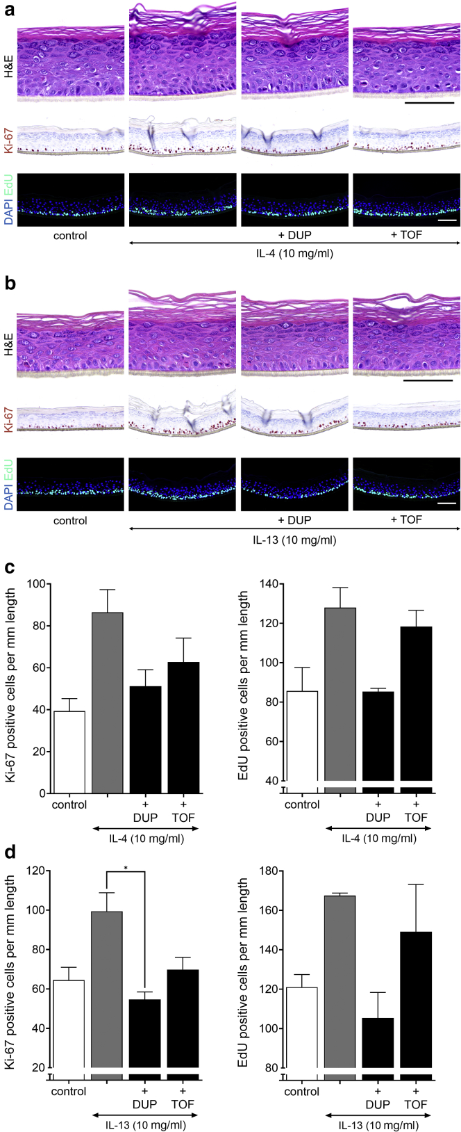
Psoriasis and atopic dermatitis are chronic inflammatory skin diseases characterized by keratinocyte (KC) hyperproliferation and epidermal acanthosis (hyperplasia). The milieu of disease-associated cytokines and soluble factors is considered a mitogenic factor; however, pinpointing the exact mitogens in this complex microenvironment is challenging. We employed organotypic human epidermal equivalents, faithfully mimicking native epidermal proliferation and stratification, to evaluate the proliferative effects of a broad panel of (literature-based) potential mitogens. The KC GF molecule, the T-helper 2 cytokines IL-4 and IL-13, and the psoriasis-associated cytokine IL-17A caused acanthosis by hyperplasia through a doubling in the number of proliferating KCs. In contrast, IFN-γ lowered proliferation, whereas IL-6, IL-20, IL-22, and oncostatin M induced acanthosis not by hyperproliferation but by hypertrophy. The T-helper 2‒cytokine‒mediated hyperproliferation was Jak/signal transducer and activator of transcription 3 dependent, whereas IL-17A and KC GF induced MAPK/extracellular signal‒regulated kinase kinase/extracellular signal‒regulated kinase‒dependent proliferation. This discovery that key regulators in atopic dermatitis and psoriasis are direct KC mitogens not only adds evidence to their crucial role in the pathophysiological processes but also highlights an additional therapeutic pillar for the mode of action of targeting biologicals (e.g., dupilumab) or small-molecule drugs (e.g., tofacitinib) by the normalization of KC turnover within the epidermal compartment.
银屑病和特应性皮炎是慢性炎症性皮肤病,其特征为角质形成细胞(KC)过度增殖和表皮棘层肥厚(增生)。疾病相关细胞因子和可溶性因子的环境被认为是一种促有丝分裂因子;然而,在这种复杂的微环境中确定确切的促有丝分裂原具有挑战性。我们采用了能忠实地模拟天然表皮增殖和分层的器官型人表皮替代物,来评估一系列(基于文献的)潜在促有丝分裂原的增殖作用。KC生长因子(GF)分子、辅助性T细胞2细胞因子白细胞介素(IL)-4和IL-13,以及银屑病相关细胞因子IL-17A,通过使增殖的KC数量翻倍,导致增生性棘层肥厚。相比之下,γ干扰素降低了增殖,而IL-6、IL-20、IL-22和制瘤素M诱导棘层肥厚不是通过过度增殖,而是通过肥大。辅助性T细胞2细胞因子介导的过度增殖依赖于Janus激酶/信号转导子和转录激活子3,而IL-17A和KC GF诱导的增殖依赖于丝裂原活化蛋白激酶/细胞外信号调节激酶激酶/细胞外信号调节激酶。这一发现,即特应性皮炎和银屑病中的关键调节因子是直接的KC促有丝分裂原,不仅为它们在病理生理过程中的关键作用增添了证据,也突出了通过使表皮层内的KC周转正常化来靶向生物制剂(如度普利尤单抗)或小分子药物(如托法替布)作用模式的另一个治疗支柱。