• 文献检索
  • 文档翻译
  • 深度研究
  • 学术资讯
  • Suppr Zotero 插件Zotero 插件
  • 邀请有礼
  • 套餐&价格
  • 历史记录
应用&插件
Suppr Zotero 插件Zotero 插件浏览器插件Mac 客户端Windows 客户端微信小程序
定价
高级版会员购买积分包购买API积分包
服务
文献检索文档翻译深度研究API 文档MCP 服务
关于我们
关于 Suppr公司介绍联系我们用户协议隐私条款
关注我们

Suppr 超能文献

核心技术专利:CN118964589B侵权必究
粤ICP备2023148730 号-1Suppr @ 2026

文献检索

告别复杂PubMed语法,用中文像聊天一样搜索,搜遍4000万医学文献。AI智能推荐,让科研检索更轻松。

立即免费搜索

文件翻译

保留排版,准确专业,支持PDF/Word/PPT等文件格式,支持 12+语言互译。

免费翻译文档

深度研究

AI帮你快速写综述,25分钟生成高质量综述,智能提取关键信息,辅助科研写作。

立即免费体验

磁共振成像用于左心室肥厚的鉴别诊断:诊断及预后意义

Magnetic Resonance for Differential Diagnosis of Left Ventricular Hypertrophy: Diagnostic and Prognostic Implications.

作者信息

Aquaro Giovanni Donato, Corsi Elisabetta, Todiere Giancarlo, Grigoratos Crysanthos, Barison Andrea, Barra Valerio, Di Bella Gianluca, Emdin Michele, Ricci Fabrizio, Pingitore Alessandro

机构信息

Fondazione Toscana G. Monasterio, 56124 Pisa, Italy.

Department of Cardiac and Thoracic medicine, Università degli studi di Pisa, 56126 Pisa, Italy.

出版信息

J Clin Med. 2022 Jan 27;11(3):651. doi: 10.3390/jcm11030651.

DOI:10.3390/jcm11030651
PMID:35160102
原文链接:https://pmc.ncbi.nlm.nih.gov/articles/PMC8836982/
Abstract

BACKGROUND

Left ventricular hypertrophy (LVH) may be due to different causes, ranging from benign secondary forms to severe cardiomyopathies. Transthoracic Echocardiography (TTE) and ECG are the first-level examinations for LVH diagnosis. Cardiac magnetic resonance (CMR) accurately defines LVH type, extent and severity.

OBJECTIVES

to evaluate the diagnostic and prognostic role of CMR in patients with TTE and/or ECG evidence of LVH.

METHODS

We performed CMR in 300 consecutive patients with echocardiographic and/or ECG signs of LVH.

RESULTS

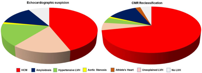
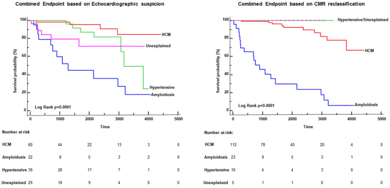
Overall, 275 patients had TTE evidence of LVH, with initial suspicion of hypertrophic cardiomyopathy (HCM) in 132 (44%), cardiac amyloidosis in 41 (14%), hypertensive LVH in 48 (16%), aortic stenosis in 4 (1%), and undetermined LVH in 50 (16%). The initial echocardiographic diagnostic suspicion of LVH was confirmed in 172 patients (57.3%) and changed in 128 patients (42.7%, < 0.0001): the diagnosis of HCM increased from 44% to 71% of patients; hypertensive and undetermined LVH decreased significantly (respectively to 4% and 5%). CMR allowed for a diagnosis in 41 out of 50 (82%) patients with undetermined LVH at TTE. CMR also identified HCM in 17 out of 25 patients with apparently normal echocardiography but with ECG criteria for LVH. Finally, the reclassification of the diagnosis by CMR was associated with a change in survival risk of patients: after CMR reclassification, no events occurred in patients with undetermined or hypertensive LVH.

CONCLUSIONS

CMR changed echocardiographic suspicion in almost half of patients with LVH. In the subgroup of patients with abnormal ECG, CMR identified LVH (particularly HCM) in 80% of patients. This study highlights the indication of CMR to better characterize the type, extent and severity of LVH detected at echocardiography and suspected with ECG.

摘要

背景

左心室肥厚(LVH)可能由多种不同原因引起,从良性继发性形式到严重的心肌病。经胸超声心动图(TTE)和心电图(ECG)是LVH诊断的一级检查方法。心脏磁共振成像(CMR)能准确界定LVH的类型、范围和严重程度。

目的

评估CMR在有TTE和/或ECG提示LVH的患者中的诊断和预后作用。

方法

我们对300例连续的有超声心动图和/或ECG显示LVH迹象的患者进行了CMR检查。

结果

总体而言,275例患者有TTE提示的LVH,最初怀疑为肥厚型心肌病(HCM)的有132例(44%),心脏淀粉样变性的有41例(14%),高血压性LVH的有48例(16%),主动脉瓣狭窄的有4例(1%),不明原因LVH的有50例(16%)。最初超声心动图对LVH的诊断怀疑在172例患者(57.3%)中得到证实,在128例患者(42.7%,P<0.0001)中发生了改变:HCM的诊断从患者的44%增加到71%;高血压性和不明原因的LVH显著减少(分别降至4%和5%)。CMR使TTE检查中50例不明原因LVH患者中的41例(82%)得以确诊。CMR还在25例超声心动图看似正常但有ECG提示LVH标准的患者中发现了17例HCM。最后,CMR对诊断的重新分类与患者生存风险的改变相关:CMR重新分类后,不明原因或高血压性LVH患者未发生事件。

结论

CMR改变了近一半LVH患者的超声心动图怀疑诊断。在ECG异常的患者亚组中,CMR在80%的患者中识别出LVH(尤其是HCM)。本研究强调了CMR在更好地界定超声心动图检测到并经ECG怀疑的LVH的类型、范围和严重程度方面的应用价值。

https://cdn.ncbi.nlm.nih.gov/pmc/blobs/0ad4/8836982/37b421c5f4dc/jcm-11-00651-g004.jpg
https://cdn.ncbi.nlm.nih.gov/pmc/blobs/0ad4/8836982/7a9f8f205f94/jcm-11-00651-g001.jpg
https://cdn.ncbi.nlm.nih.gov/pmc/blobs/0ad4/8836982/bdb4cd7808b0/jcm-11-00651-g002.jpg
https://cdn.ncbi.nlm.nih.gov/pmc/blobs/0ad4/8836982/9e5032d695ae/jcm-11-00651-g003.jpg
https://cdn.ncbi.nlm.nih.gov/pmc/blobs/0ad4/8836982/37b421c5f4dc/jcm-11-00651-g004.jpg

相似文献

1
Magnetic Resonance for Differential Diagnosis of Left Ventricular Hypertrophy: Diagnostic and Prognostic Implications.磁共振成像用于左心室肥厚的鉴别诊断:诊断及预后意义
J Clin Med. 2022 Jan 27;11(3):651. doi: 10.3390/jcm11030651.
2
Discrepancies in Left Ventricular Mass Calculation Based on Echocardiography and Cardiovascular Magnetic Resonance Measurements in Patients with Left Ventricular Hypertrophy.基于超声心动图和心血管磁共振测量的左心室肥厚患者左心室质量计算中的差异。
J Am Soc Echocardiogr. 2015 Oct;28(10):1194-1203, e2. doi: 10.1016/j.echo.2015.06.009. Epub 2015 Jul 17.
3
Contemporary family screening in hypertrophic cardiomyopathy: the role of cardiovascular magnetic resonance.肥厚型心肌病的现代家族筛查:心血管磁共振的作用。
Eur Heart J Cardiovasc Imaging. 2022 Aug 22;23(9):1144-1154. doi: 10.1093/ehjci/jeac099.
4
Comparison of echocardiographic and cardiac magnetic resonance imaging in hypertrophic cardiomyopathy sarcomere mutation carriers without left ventricular hypertrophy.肥厚型心肌病肌节突变携带者无左心室肥厚时超声心动图与心脏磁共振成像的比较
Circ Cardiovasc Genet. 2013 Jun;6(3):230-7. doi: 10.1161/CIRCGENETICS.113.000037. Epub 2013 May 20.
5
Intelligent diagnosis of left ventricular hypertrophy using transthoracic echocardiography videos.经胸超声心动图视频左心室肥厚的智能诊断。
Comput Methods Programs Biomed. 2022 Nov;226:107182. doi: 10.1016/j.cmpb.2022.107182. Epub 2022 Oct 12.
6
Ongoing assertion of two-dimensional measurements on differentiation type of left ventricular hypertrophy: Focus on inferior vena cava.持续对左心室肥厚的分化类型进行二维测量:重点关注下腔静脉。
Echocardiography. 2024 Jul;41(7):e15880. doi: 10.1111/echo.15880.
7
Distinct pressure half-time values by transthoracic echocardiography for grading of paravalvular regurgitation after transcatheter aortic valve replacement.经胸超声心动图测量瓣周漏反流压差减半时间用于分级评估经导管主动脉瓣置换术后瓣周漏
Sci Rep. 2020 Feb 13;10(1):2549. doi: 10.1038/s41598-020-59211-z.
8
Echocardiographic strain in hypertrophic cardiomyopathy and hypertensive left ventricular hypertrophy.肥厚型心肌病和高血压性左心室肥厚中的超声心动图应变
Echocardiography. 2019 Feb;36(2):257-265. doi: 10.1111/echo.14222. Epub 2018 Dec 18.
9
Association of electrocardiographic and imaging surrogates of left ventricular hypertrophy with incident atrial fibrillation: MESA (Multi-Ethnic Study of Atherosclerosis).左心室肥厚的心电图和影像学指标与心房颤动事件的相关性:MESA(动脉粥样硬化多民族研究)。
J Am Coll Cardiol. 2014 May 20;63(19):2007-13. doi: 10.1016/j.jacc.2014.01.066. Epub 2014 Mar 19.
10
Electrocardiographic criteria for left ventricular hypertrophy in aortic valve stenosis: Correlation with echocardiographic parameters.主动脉瓣狭窄时左心室肥厚的心电图标准:与超声心动图参数的相关性。
Ann Noninvasive Electrocardiol. 2019 Sep;24(5):e12645. doi: 10.1111/anec.12645. Epub 2019 Mar 21.

引用本文的文献

1
The Depiction of Hypertension in Heart Imaging Examinations: An Up-to-Date Review of the Evidence.心脏影像学检查中高血压的描述:证据的最新综述。
Vasc Health Risk Manag. 2023 Nov 28;19:789-796. doi: 10.2147/VHRM.S436133. eCollection 2023.
2
Electrocardiographic Parameters Associated with Adverse Outcomes in Children with Cardiomyopathies.与心肌病患儿不良结局相关的心电图参数
J Clin Med. 2022 Nov 24;11(23):6930. doi: 10.3390/jcm11236930.
3
Cardiac Magnetic Resonance in Fabry Disease: Morphological, Functional, and Tissue Features.

本文引用的文献

1
Cardiovascular Magnetic Resonance for the Differentiation of Left Ventricular Hypertrophy.心血管磁共振在左心室肥厚鉴别诊断中的应用。
Curr Heart Fail Rep. 2020 Oct;17(5):192-204. doi: 10.1007/s11897-020-00481-z.
2
Standardized cardiovascular magnetic resonance imaging (CMR) protocols: 2020 update.标准化心血管磁共振成像(CMR)协议:2020 年更新。
J Cardiovasc Magn Reson. 2020 Feb 24;22(1):17. doi: 10.1186/s12968-020-00607-1.
3
Myocardial Tissue Characterization and Fibrosis by Imaging.心肌组织的影像学特征与纤维化
法布里病的心脏磁共振成像:形态学、功能及组织特征
Diagnostics (Basel). 2022 Nov 1;12(11):2652. doi: 10.3390/diagnostics12112652.
4
Post-Mortem Cardiac Magnetic Resonance in Explanted Heart of Patients with Sudden Death.心脏移植术后猝死患者的心脏磁共振成像。
Int J Environ Res Public Health. 2022 Oct 17;19(20):13395. doi: 10.3390/ijerph192013395.
5
Usefulness of Longitudinal Strain Adjusted to Regional Thickness in Hypertrophic Cardiomyopathy.肥厚型心肌病中经区域厚度调整的纵向应变的效用。
J Clin Med. 2022 Apr 8;11(8):2089. doi: 10.3390/jcm11082089.
JACC Cardiovasc Imaging. 2020 May;13(5):1221-1234. doi: 10.1016/j.jcmg.2019.06.030. Epub 2019 Sep 18.
4
Prevalence of Unexplained Left Ventricular Hypertrophy by Cardiac Magnetic Resonance Imaging in MESA.磁共振成像在 MESA 研究中不明原因左心室肥厚的患病率。
J Am Heart Assoc. 2019 Apr 16;8(8):e012250. doi: 10.1161/JAHA.119.012250.
5
2018 ESC/ESH Guidelines for the management of arterial hypertension.2018年欧洲心脏病学会/欧洲高血压学会动脉高血压管理指南。
Eur Heart J. 2018 Sep 1;39(33):3021-3104. doi: 10.1093/eurheartj/ehy339.
6
2017 ESC/EACTS Guidelines for the management of valvular heart disease.2017年欧洲心脏病学会/欧洲心胸外科学会瓣膜性心脏病管理指南。
Eur Heart J. 2017 Sep 21;38(36):2739-2791. doi: 10.1093/eurheartj/ehx391.
7
Discrepant Measurements of Maximal Left Ventricular Wall Thickness Between Cardiac Magnetic Resonance Imaging and Echocardiography in Patients With Hypertrophic Cardiomyopathy.肥厚型心肌病患者心脏磁共振成像与超声心动图测量左心室最大壁厚的差异
Circ Cardiovasc Imaging. 2017 Aug;10(8). doi: 10.1161/CIRCIMAGING.117.006309.
8
Comparison of Maximal Wall Thickness in Hypertrophic Cardiomyopathy Differs Between Magnetic Resonance Imaging and Transthoracic Echocardiography.肥厚型心肌病最大室壁厚度在磁共振成像和经胸超声心动图检查中的比较存在差异。
Am J Cardiol. 2017 Feb 15;119(4):643-650. doi: 10.1016/j.amjcard.2016.11.010. Epub 2016 Nov 16.
9
Reference values of cardiac volumes, dimensions, and new functional parameters by MR: A multicenter, multivendor study.磁共振成像测定心脏容积、尺寸及新功能参数的参考值:一项多中心、多厂商研究
J Magn Reson Imaging. 2017 Apr;45(4):1055-1067. doi: 10.1002/jmri.25450. Epub 2016 Aug 29.
10
Prognostic Value of LGE-CMR in HCM: A Meta-Analysis.LGE-CMR 在 HCM 中的预后价值:一项荟萃分析。
JACC Cardiovasc Imaging. 2016 Dec;9(12):1392-1402. doi: 10.1016/j.jcmg.2016.02.031. Epub 2016 Jul 20.