• 文献检索
  • 文档翻译
  • 深度研究
  • 学术资讯
  • Suppr Zotero 插件Zotero 插件
  • 邀请有礼
  • 套餐&价格
  • 历史记录
应用&插件
Suppr Zotero 插件Zotero 插件浏览器插件Mac 客户端Windows 客户端微信小程序
定价
高级版会员购买积分包购买API积分包
服务
文献检索文档翻译深度研究API 文档MCP 服务
关于我们
关于 Suppr公司介绍联系我们用户协议隐私条款
关注我们

Suppr 超能文献

核心技术专利:CN118964589B侵权必究
粤ICP备2023148730 号-1Suppr @ 2026

文献检索

告别复杂PubMed语法,用中文像聊天一样搜索,搜遍4000万医学文献。AI智能推荐,让科研检索更轻松。

立即免费搜索

文件翻译

保留排版,准确专业,支持PDF/Word/PPT等文件格式,支持 12+语言互译。

免费翻译文档

深度研究

AI帮你快速写综述,25分钟生成高质量综述,智能提取关键信息,辅助科研写作。

立即免费体验

缺氧诱导因子-1 的抑制导致糖尿病中线粒体活性氧的产生增加。

Repression of hypoxia-inducible factor-1 contributes to increased mitochondrial reactive oxygen species production in diabetes.

机构信息

Department of Molecular Medicine and Surgery, Karolinska Institutet, Stockholm, Sweden.

Centre for Inherited Cardiovascular Diseases, IRCCS Foundation University Hospital Policlinico San Matteo, Pavia, Italy.

出版信息

Elife. 2022 Feb 15;11:e70714. doi: 10.7554/eLife.70714.

DOI:10.7554/eLife.70714
PMID:35164902
原文链接:https://pmc.ncbi.nlm.nih.gov/articles/PMC8846593/
Abstract

BACKGROUND

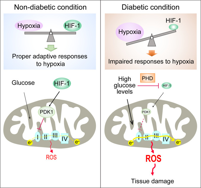
Excessive production of mitochondrial reactive oxygen species (ROS) is a central mechanism for the development of diabetes complications. Recently, hypoxia has been identified to play an additional pathogenic role in diabetes. In this study, we hypothesized that ROS overproduction was secondary to the impaired responses to hypoxia due to the inhibition of hypoxia-inducible factor-1 (HIF-1) by hyperglycemia.

METHODS

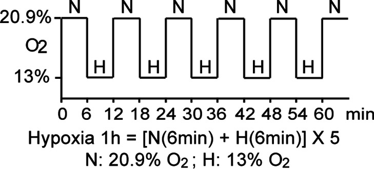
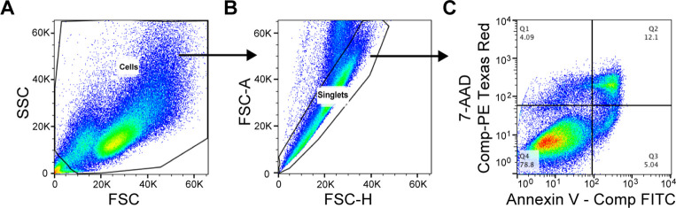
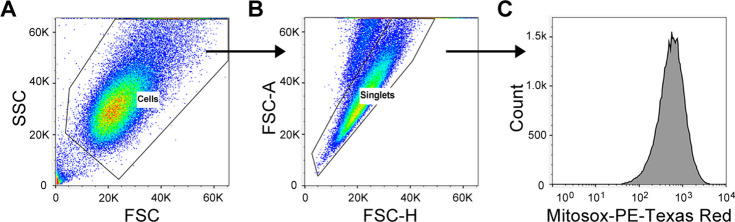
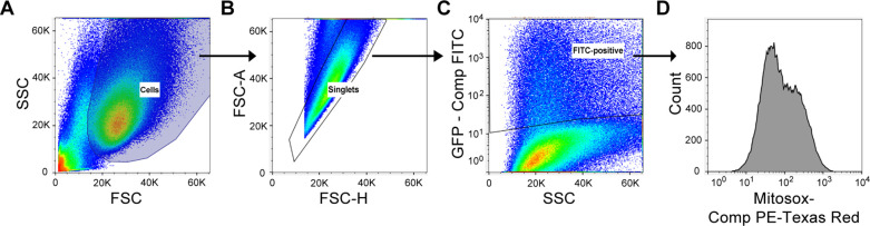
The ROS levels were analyzed in the blood of healthy subjects and individuals with type 1 diabetes after exposure to hypoxia. The relation between HIF-1, glucose levels, ROS production and its functional consequences were analyzed in renal mIMCD-3 cells and in kidneys of mouse models of diabetes.

RESULTS

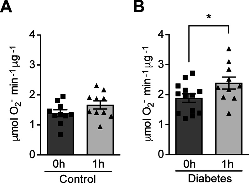
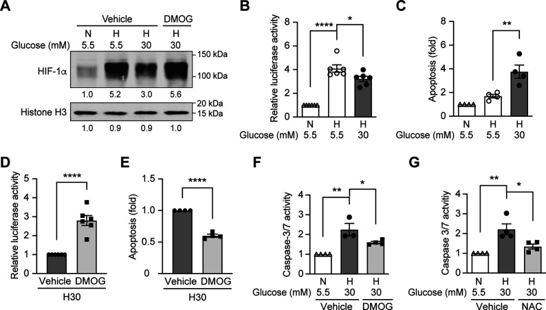
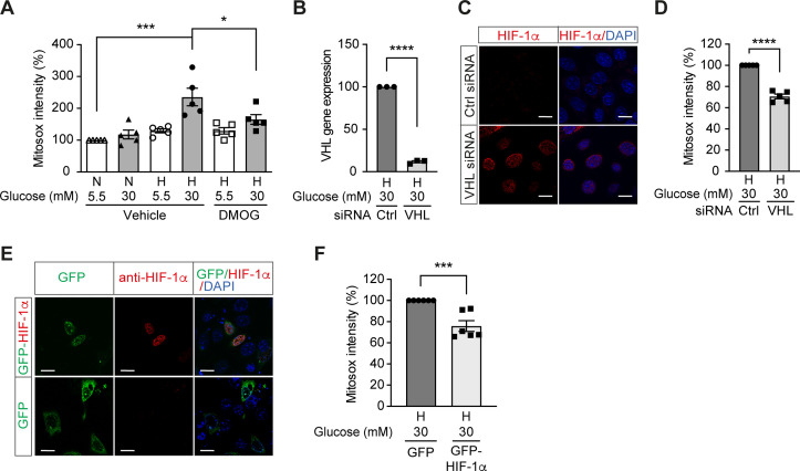
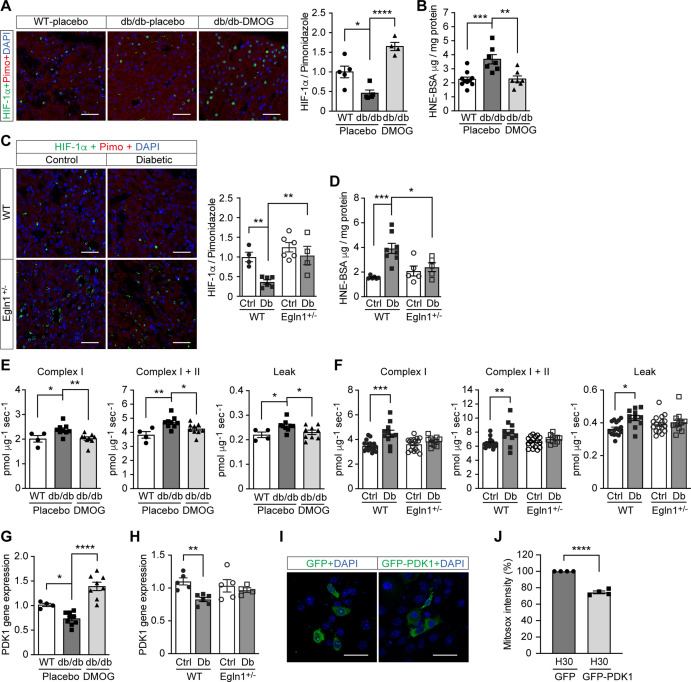
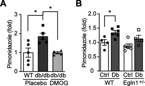
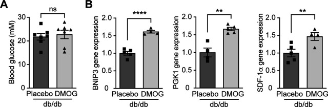
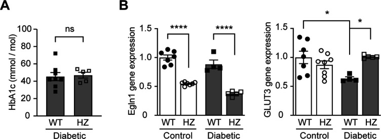
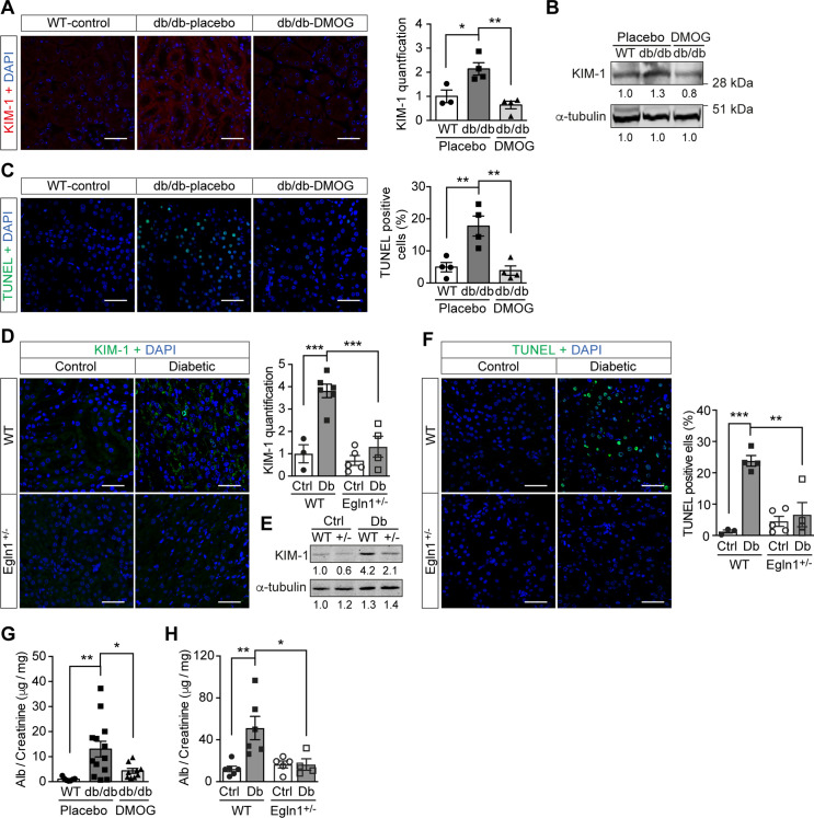
Exposure to hypoxia increased circulating ROS in subjects with diabetes, but not in subjects without diabetes. High glucose concentrations repressed HIF-1 both in hypoxic cells and in kidneys of animals with diabetes, through a HIF prolyl-hydroxylase (PHD)-dependent mechanism. The impaired HIF-1 signaling contributed to excess production of mitochondrial ROS through increased mitochondrial respiration that was mediated by Pyruvate dehydrogenase kinase 1 (PDK1). The restoration of HIF-1 function attenuated ROS overproduction despite persistent hyperglycemia, and conferred protection against apoptosis and renal injury in diabetes.

CONCLUSIONS

We conclude that the repression of HIF-1 plays a central role in mitochondrial ROS overproduction in diabetes and is a potential therapeutic target for diabetic complications. These findings are timely since the first PHD inhibitor that can activate HIF-1 has been newly approved for clinical use.

FUNDING

This work was supported by grants from the Swedish Research Council, Stockholm County Research Council, Stockholm Regional Research Foundation, Bert von Kantzows Foundation, Swedish Society of Medicine, Kung Gustaf V:s och Drottning Victorias Frimurarestifelse, Karolinska Institute's Research Foundations, Strategic Research Programme in Diabetes, and Erling-Persson Family Foundation for S-B.C.; grants from the Swedish Research Council and Swedish Heart and Lung Foundation for T.A.S.; and ERC consolidator grant for M.M.

摘要

背景

线粒体活性氧(ROS)的过度产生是糖尿病并发症发展的核心机制。最近,缺氧被确定在糖尿病中发挥额外的致病作用。在这项研究中,我们假设 ROS 的过度产生是由于高血糖抑制缺氧诱导因子-1(HIF-1),导致对缺氧的反应受损所致。

方法

分析健康受试者和 1 型糖尿病患者在暴露于缺氧后血液中的 ROS 水平。在肾 mIMCD-3 细胞和糖尿病小鼠模型的肾脏中分析 HIF-1、葡萄糖水平、ROS 产生及其功能后果之间的关系。

结果

暴露于缺氧会增加糖尿病患者的循环 ROS,但不会增加非糖尿病患者的循环 ROS。高葡萄糖浓度通过依赖 HIF 脯氨酰羟化酶(PHD)的机制,在缺氧细胞和糖尿病动物的肾脏中抑制 HIF-1。受损的 HIF-1 信号通过增加介导的丙酮酸脱氢酶激酶 1(PDK1)来促进线粒体 ROS 的过度产生。尽管持续高血糖,但恢复 HIF-1 功能可减轻 ROS 过度产生,并在糖尿病中提供对细胞凋亡和肾损伤的保护作用。

结论

我们得出结论,HIF-1 的抑制在糖尿病中线粒体 ROS 的过度产生中起核心作用,是糖尿病并发症的潜在治疗靶点。这些发现是及时的,因为新批准了第一种可以激活 HIF-1 的 PHD 抑制剂用于临床使用。

资金

本工作得到瑞典研究理事会、斯德哥尔摩郡研究理事会、斯德哥尔摩地区研究基金会、Bert von Kantzows 基金会、瑞典医学协会、古斯塔夫五世和王后维多利亚基金会、卡罗林斯卡研究所研究基金会、糖尿病战略研究计划以及 Erling-Persson 家族基金会的资助;得到瑞典研究理事会和瑞典心脏和肺基金会的资助;以及 ERC 巩固者资助。

https://cdn.ncbi.nlm.nih.gov/pmc/blobs/7b06/8846593/ef0d59beb6d2/elife-70714-fig6.jpg
https://cdn.ncbi.nlm.nih.gov/pmc/blobs/7b06/8846593/516acaf28741/elife-70714-fig1.jpg
https://cdn.ncbi.nlm.nih.gov/pmc/blobs/7b06/8846593/caf14cd9f64c/elife-70714-fig1-figsupp1.jpg
https://cdn.ncbi.nlm.nih.gov/pmc/blobs/7b06/8846593/31653ae544f4/elife-70714-fig2.jpg
https://cdn.ncbi.nlm.nih.gov/pmc/blobs/7b06/8846593/422d25ffbdac/elife-70714-fig2-figsupp1.jpg
https://cdn.ncbi.nlm.nih.gov/pmc/blobs/7b06/8846593/a3e9c23504ac/elife-70714-fig3.jpg
https://cdn.ncbi.nlm.nih.gov/pmc/blobs/7b06/8846593/5930b6fab7ea/elife-70714-fig3-figsupp1.jpg
https://cdn.ncbi.nlm.nih.gov/pmc/blobs/7b06/8846593/bc4fcde0f306/elife-70714-fig3-figsupp2.jpg
https://cdn.ncbi.nlm.nih.gov/pmc/blobs/7b06/8846593/c165b0801b97/elife-70714-fig4.jpg
https://cdn.ncbi.nlm.nih.gov/pmc/blobs/7b06/8846593/b0d7c034116a/elife-70714-fig4-figsupp1.jpg
https://cdn.ncbi.nlm.nih.gov/pmc/blobs/7b06/8846593/fea892b9a0bc/elife-70714-fig4-figsupp2.jpg
https://cdn.ncbi.nlm.nih.gov/pmc/blobs/7b06/8846593/5875f7464d4d/elife-70714-fig4-figsupp3.jpg
https://cdn.ncbi.nlm.nih.gov/pmc/blobs/7b06/8846593/090878df202f/elife-70714-fig5.jpg
https://cdn.ncbi.nlm.nih.gov/pmc/blobs/7b06/8846593/ef0d59beb6d2/elife-70714-fig6.jpg

相似文献

1
Repression of hypoxia-inducible factor-1 contributes to increased mitochondrial reactive oxygen species production in diabetes.缺氧诱导因子-1 的抑制导致糖尿病中线粒体活性氧的产生增加。
Elife. 2022 Feb 15;11:e70714. doi: 10.7554/eLife.70714.
2
Hypoxia Triggers Osteochondrogenic Differentiation of Vascular Smooth Muscle Cells in an HIF-1 (Hypoxia-Inducible Factor 1)-Dependent and Reactive Oxygen Species-Dependent Manner.缺氧以依赖 HIF-1(缺氧诱导因子 1)和活性氧的方式触发血管平滑肌细胞的成骨软骨分化。
Arterioscler Thromb Vasc Biol. 2019 Jun;39(6):1088-1099. doi: 10.1161/ATVBAHA.119.312509.
3
HIF-1 prevents the overproduction of mitochondrial ROS after cytokine stimulation through induction of PDK-1.缺氧诱导因子-1通过诱导丙酮酸脱氢酶激酶-1,防止细胞因子刺激后线粒体活性氧的过度产生。
Cell Cycle. 2009 Sep 1;8(17):2844-9. doi: 10.4161/cc.8.17.9544. Epub 2009 Sep 16.
4
mtDNA as a Mediator for Expression of Hypoxia-Inducible Factor 1α and ROS in Hypoxic Neuroblastoma Cells.线粒体DNA作为缺氧神经母细胞瘤细胞中缺氧诱导因子1α和活性氧表达的介质
Int J Mol Sci. 2017 Jun 7;18(6):1220. doi: 10.3390/ijms18061220.
5
Hypoxia-inducible factor-1 activation in nonhypoxic conditions: the essential role of mitochondrial-derived reactive oxygen species.缺氧诱导因子-1 在非缺氧条件下的激活:线粒体来源的活性氧的重要作用。
Mol Biol Cell. 2010 Sep 15;21(18):3247-57. doi: 10.1091/mbc.E10-01-0025. Epub 2010 Jul 21.
6
Pyruvate dehydrogenase kinase 1 protects against neuronal injury and memory loss in mouse models of diabetes.丙酮酸脱氢酶激酶 1 可保护糖尿病小鼠模型免受神经元损伤和记忆丧失。
Cell Death Dis. 2023 Nov 7;14(11):722. doi: 10.1038/s41419-023-06249-2.
7
Mitochondrial reactive oxygen species trigger hypoxia-inducible factor-dependent extension of the replicative life span during hypoxia.线粒体活性氧在缺氧过程中触发缺氧诱导因子依赖性的复制寿命延长。
Mol Cell Biol. 2007 Aug;27(16):5737-45. doi: 10.1128/MCB.02265-06. Epub 2007 Jun 11.
8
Bicalutamide Elicits Renal Damage by Causing Mitochondrial Dysfunction ROS Damage and Upregulation of HIF-1.比卡鲁胺通过引起线粒体功能障碍、活性氧损伤和缺氧诱导因子-1上调引发肾损伤。
Int J Mol Sci. 2020 May 11;21(9):3400. doi: 10.3390/ijms21093400.
9
Reactive Oxygen Species Formation in the Brain at Different Oxygen Levels: The Role of Hypoxia Inducible Factors.不同氧水平下大脑中活性氧的形成:缺氧诱导因子的作用
Front Cell Dev Biol. 2018 Oct 10;6:132. doi: 10.3389/fcell.2018.00132. eCollection 2018.
10
Hypoxia-inducible factor 1: regulator of mitochondrial metabolism and mediator of ischemic preconditioning.缺氧诱导因子1:线粒体代谢的调节因子和缺血预处理的介质。
Biochim Biophys Acta. 2011 Jul;1813(7):1263-8. doi: 10.1016/j.bbamcr.2010.08.006. Epub 2010 Aug 21.

引用本文的文献

1
Mitochondria in oxidative stress, inflammation and aging: from mechanisms to therapeutic advances.氧化应激、炎症与衰老中的线粒体:从机制到治疗进展
Signal Transduct Target Ther. 2025 Jun 11;10(1):190. doi: 10.1038/s41392-025-02253-4.
2
Human Adipose Tissue Metabolism in Obesity.肥胖状态下的人体脂肪组织代谢
J Obes Metab Syndr. 2025 Apr 30;34(2):105-119. doi: 10.7570/jomes25025. Epub 2025 Apr 8.
3
Angiogenesis during diabetic wound repair: from mechanism to therapy opportunity.糖尿病伤口修复过程中的血管生成:从机制到治疗机会

本文引用的文献

1
Hypoxia and hypoxia-inducible factors in diabetes and its complications.糖尿病及其并发症中的缺氧和缺氧诱导因子。
Diabetologia. 2021 Apr;64(4):709-716. doi: 10.1007/s00125-021-05380-z. Epub 2021 Jan 26.
2
Oxidative Stress and Inflammation in Renal and Cardiovascular Complications of Diabetes.糖尿病肾脏及心血管并发症中的氧化应激与炎症
Biology (Basel). 2020 Dec 30;10(1):18. doi: 10.3390/biology10010018.
3
Na controls hypoxic signalling by the mitochondrial respiratory chain.钠通过线粒体呼吸链控制低氧信号。
Burns Trauma. 2025 Feb 7;13:tkae052. doi: 10.1093/burnst/tkae052. eCollection 2025.
4
Roxadustat improves diabetic myocardial injury by upregulating HIF-1α/UCP2 against oxidative stress.罗沙司他通过上调缺氧诱导因子-1α/解偶联蛋白2对抗氧化应激来改善糖尿病性心肌损伤。
Cardiovasc Diabetol. 2025 Feb 7;24(1):67. doi: 10.1186/s12933-025-02601-2.
5
Hypoxia Regulates Brown Adipocyte Differentiation and Stimulates miR-210 by HIF-1α.缺氧通过HIF-1α调节棕色脂肪细胞分化并刺激miR-210。
Int J Mol Sci. 2024 Dec 26;26(1):117. doi: 10.3390/ijms26010117.
6
Advances in Smart-Response Hydrogels for Skin Wound Repair.用于皮肤伤口修复的智能响应水凝胶研究进展
Polymers (Basel). 2024 Oct 5;16(19):2818. doi: 10.3390/polym16192818.
7
Roles of β-Cell Hypoxia in the Progression of Type 2 Diabetes.β细胞缺氧在2型糖尿病进展中的作用
Int J Mol Sci. 2024 Apr 10;25(8):4186. doi: 10.3390/ijms25084186.
8
Halofuginone prevents outer retinal degeneration in a mouse model of light-induced retinopathy.哈尔福林酮可预防光诱导性视网膜病变小鼠模型的视网膜外层变性。
PLoS One. 2024 Mar 27;19(3):e0300045. doi: 10.1371/journal.pone.0300045. eCollection 2024.
9
Appropriate glycemic management protects the germline but not the uterine environment in hyperglycemia.适当的血糖管理可以保护生殖细胞,但不能保护高血糖中的子宫环境。
EMBO Rep. 2024 Apr;25(4):1752-1772. doi: 10.1038/s44319-024-00097-7. Epub 2024 Mar 15.
10
SKP alleviates the ferroptosis in diabetic kidney disease through suppression of HIF-1α/HO-1 pathway based on network pharmacology analysis and experimental validation.基于网络药理学分析和实验验证,SKP通过抑制HIF-1α/HO-1通路减轻糖尿病肾病中的铁死亡。
Chin Med. 2024 Feb 25;19(1):31. doi: 10.1186/s13020-024-00901-5.
Nature. 2020 Oct;586(7828):287-291. doi: 10.1038/s41586-020-2551-y. Epub 2020 Jul 29.
4
The role of oxidative stress and hypoxia in renal disease.氧化应激和缺氧在肾脏疾病中的作用。
Kidney Res Clin Pract. 2019 Dec 31;38(4):414-426. doi: 10.23876/j.krcp.19.063.
5
Mechanisms of hypoxia signalling: new implications for nephrology.缺氧信号转导机制:对肾脏病学的新启示。
Nat Rev Nephrol. 2019 Oct;15(10):641-659. doi: 10.1038/s41581-019-0182-z. Epub 2019 Sep 5.
6
Roxadustat for Anemia in Patients with Kidney Disease Not Receiving Dialysis.罗沙司他治疗未接受透析的肾病患者的贫血。
N Engl J Med. 2019 Sep 12;381(11):1001-1010. doi: 10.1056/NEJMoa1813599. Epub 2019 Jul 24.
7
Kidney outer medulla mitochondria are more efficient compared with cortex mitochondria as a strategy to sustain ATP production in a suboptimal environment.肾脏外髓质的线粒体与皮质线粒体相比效率更高,这是一种在环境不佳的情况下维持 ATP 产生的策略。
Am J Physiol Renal Physiol. 2018 Sep 1;315(3):F677-F681. doi: 10.1152/ajprenal.00207.2018. Epub 2018 May 30.
8
Deletion of Uncoupling Protein-2 reduces renal mitochondrial leak respiration, intrarenal hypoxia and proteinuria in a mouse model of type 1 diabetes.解偶联蛋白-2 的缺失可减少 1 型糖尿病小鼠模型的肾线粒体渗漏呼吸、肾内缺氧和蛋白尿。
Acta Physiol (Oxf). 2018 Aug;223(4):e13058. doi: 10.1111/apha.13058. Epub 2018 Mar 15.
9
Electron Paramagnetic Resonance Measurements of Reactive Oxygen Species by Cyclic Hydroxylamine Spin Probes.循环羟胺自旋探针的活性氧的电子顺磁共振测量。
Antioxid Redox Signal. 2018 May 20;28(15):1433-1443. doi: 10.1089/ars.2017.7396. Epub 2017 Nov 17.
10
Oxygen-induced impairment in arterial function is corrected by slow breathing in patients with type 1 diabetes.吸氧导致的 1 型糖尿病患者动脉功能障碍可通过缓慢呼吸得到纠正。
Sci Rep. 2017 Jul 20;7(1):6001. doi: 10.1038/s41598-017-04947-4.