Institute of Neuropathology, University Hospital of Zurich, Zurich, Switzerland.
Amyloidosis Research and Treatment Center, Foundation Scientific Institute Policlinico San Matteo, Pavia, Italy.
Brain Pathol. 2022 Sep;32(5):e13056. doi: 10.1111/bpa.13056. Epub 2022 Feb 17.
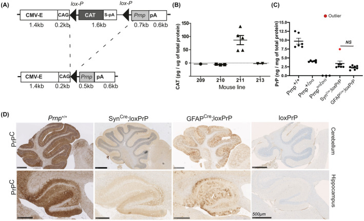
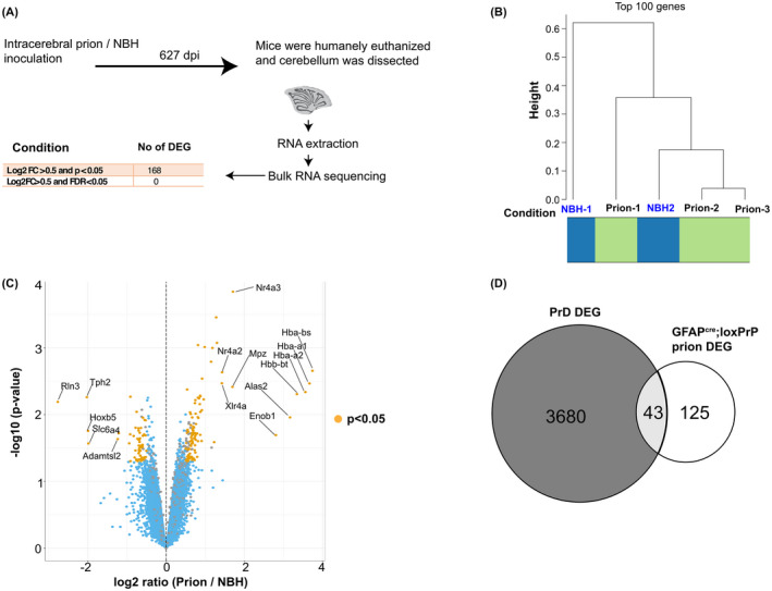
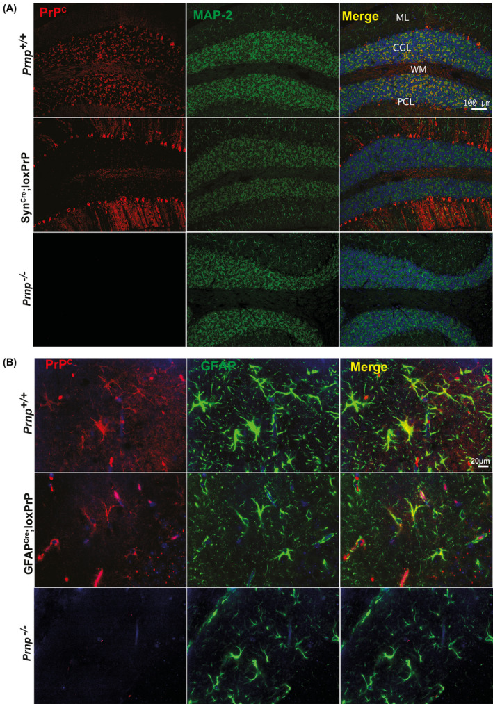
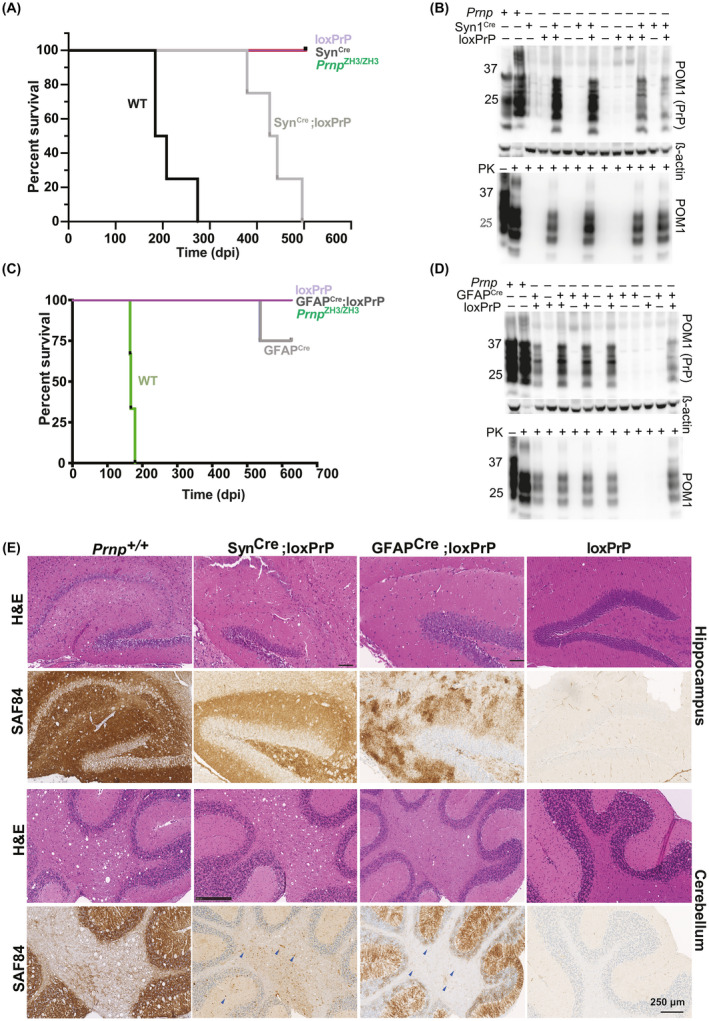
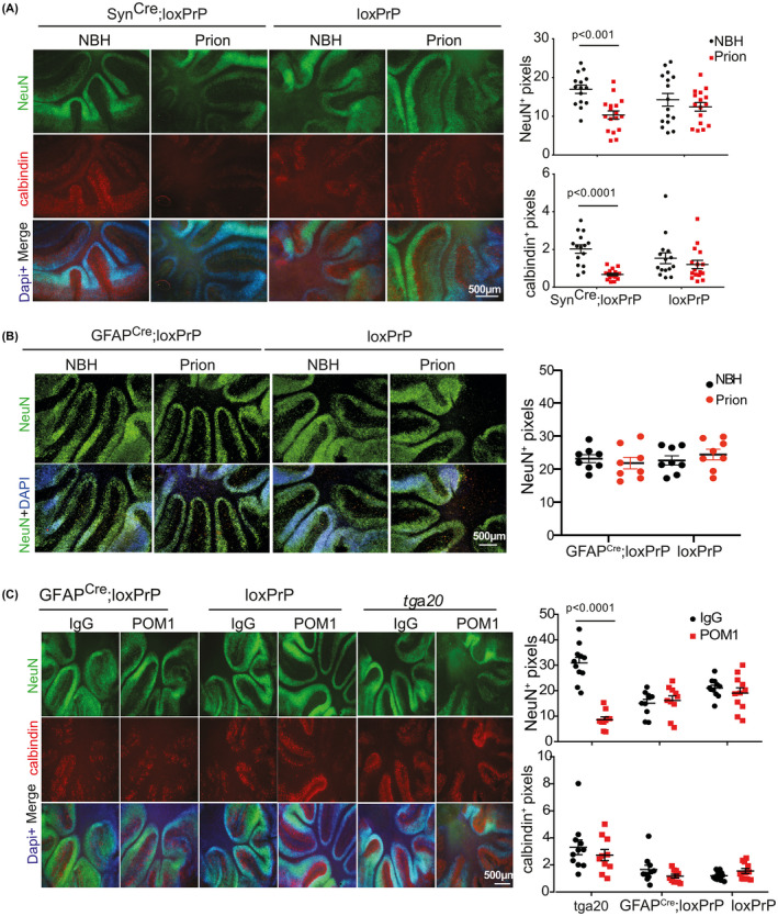
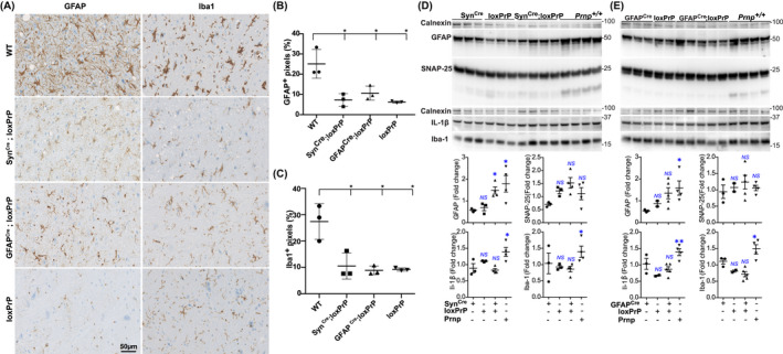
Although prion infections cause cognitive impairment and neuronal death, transcriptional and translational profiling shows progressive derangement within glia but surprisingly little changes within neurons. Here we expressed PrP selectively in neurons and astrocytes of mice. After prion infection, both astrocyte and neuron-restricted PrP expression led to copious brain accumulation of PrP . As expected, neuron-restricted expression was associated with typical prion disease. However, mice with astrocyte-restricted PrP expression experienced a normal life span, did not develop clinical disease, and did not show astro- or microgliosis. Besides confirming that PrP is innocuous to PrP -deficient neurons, these results show that astrocyte-born PrP does not activate the extreme neuroinflammation that accompanies the onset of prion disease and precedes any molecular changes of neurons. This points to a nonautonomous mechanism by which prion-infected neurons instruct astrocytes and microglia to acquire a specific cellular state that, in turn, drives neural dysfunction.
尽管朊病毒感染会导致认知障碍和神经元死亡,但转录和翻译谱分析显示,神经胶质细胞内的功能紊乱呈进行性发展,而神经元内的变化却出人意料地少。在这里,我们在小鼠的神经元和星形胶质细胞中选择性地表达了 PrP。在朊病毒感染后,星形胶质细胞和神经元特异性表达 PrP 均导致大量 PrP 在脑中积累。正如预期的那样,神经元特异性表达与典型的朊病毒病有关。然而,星形胶质细胞特异性表达 PrP 的小鼠具有正常的寿命,不会发展为临床疾病,也不会出现星形胶质细胞或小胶质细胞增生。除了证实 PrP 对 PrP 缺陷神经元是无害的之外,这些结果表明,星形胶质细胞产生的 PrP 不会激活伴随朊病毒病发作并先于神经元任何分子变化出现的极度神经炎症。这表明,朊病毒感染的神经元通过非自主机制指示星形胶质细胞和小胶质细胞获得特定的细胞状态,进而导致神经功能障碍。