Radiology Department, Medical Park University, Istanbul 34262, Turkey.
Radiology Department, Oncology Research Hospital, University of Health Sciences, Ankara 06610, Turkey.
Medicina (Kaunas). 2022 Feb 1;58(2):221. doi: 10.3390/medicina58020221.
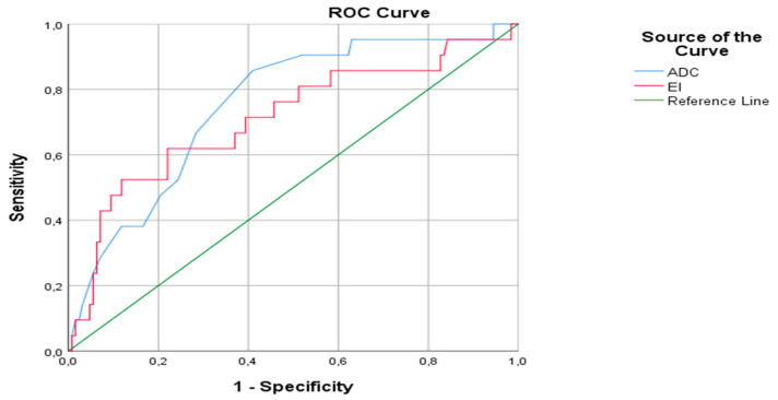
: Renal Cell Carcinoma (RCC) accounts for 85% and oncocytomas constitute 3-7% of solid renal masses. Oncocytomas can be confused, especially with hypovascular RCC. The purpose of this research was to evaluate the contribution of diffusion-weighted imaging (DWI) and contrast-enhanced MRI sequences in the differential diagnosis of RCC and oncocytoma : 465 patients with the diagnosis of RCC and 45 patients diagnosed with oncocytoma were retrospectively reviewed between 2009 to 2020. All MRI acquisitions were handled by a 1.5 T device (Achieva, Philips Healthcare, Best, The Netherlands) and all images were evaluated by the consensus of two radiologists with 10-15 years' experience. The SPSS package program version 15.0 software was used for statistical analysis of the study. Chi-square test, Mann-Whitney U test or the Kruskal-Wallis tests were used in the statistical analysis. A receiver operating characteristic (ROC) curve was used to calculate the cut-off values : The results were evaluated with a 95% confidence interval and a significance threshold of < 0.05. ADC values ( < 0.001) and enhancement index ( < 0.01) were significantly lower in the RCC group than the oncocytoma group. : DWI might become an alternative technique to the contrast-enhanced MRI in patients with contrast agent nephropathy or with a high risk of nephrogenic systemic fibrosis, calculation of CI of the oncocytoma and RCCs in the contrast-enhanced acquisitions would contribute to the differential diagnosis.
肾细胞癌 (RCC) 占 85%,而嗜酸细胞瘤构成 3-7%的实体性肾肿块。嗜酸细胞瘤可能会被混淆,特别是与低血供 RCC 混淆。本研究旨在评估扩散加权成像 (DWI) 和对比增强 MRI 序列在 RCC 和嗜酸细胞瘤鉴别诊断中的作用:2009 年至 2020 年间回顾性分析了 465 例 RCC 诊断患者和 45 例嗜酸细胞瘤诊断患者。所有 MRI 采集均由 1.5T 设备 (Achieva,Philips Healthcare,Best,荷兰) 处理,所有图像均由两位具有 10-15 年经验的放射科医生共识评估。使用 SPSS 包程序版本 15.0 软件进行研究的统计分析。卡方检验、Mann-Whitney U 检验或 Kruskal-Wallis 检验用于统计分析。使用受试者工作特征 (ROC) 曲线计算截断值:使用 95%置信区间和 < 0.05 的显著性阈值评估结果。RCC 组的 ADC 值(<0.001)和增强指数(<0.01)显著低于嗜酸细胞瘤组。在造影剂肾病或存在高风险发生肾源性系统性纤维化的患者中,DWI 可能成为对比增强 MRI 的替代技术,计算对比增强采集的嗜酸细胞瘤和 RCC 的 CI 将有助于鉴别诊断。