Department of Chemical and Biological Engineering, University of Sheffield, Mappin St, Sheffield S1 3JD, UK.
Department of Oncology and Metabolism, The Medical School, University of Sheffield, Beech Hill Road, Sheffield S10 2RX, UK.
Int J Mol Sci. 2022 Feb 10;23(4):1961. doi: 10.3390/ijms23041961.
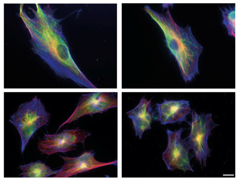
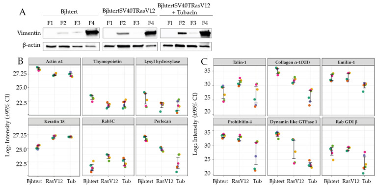
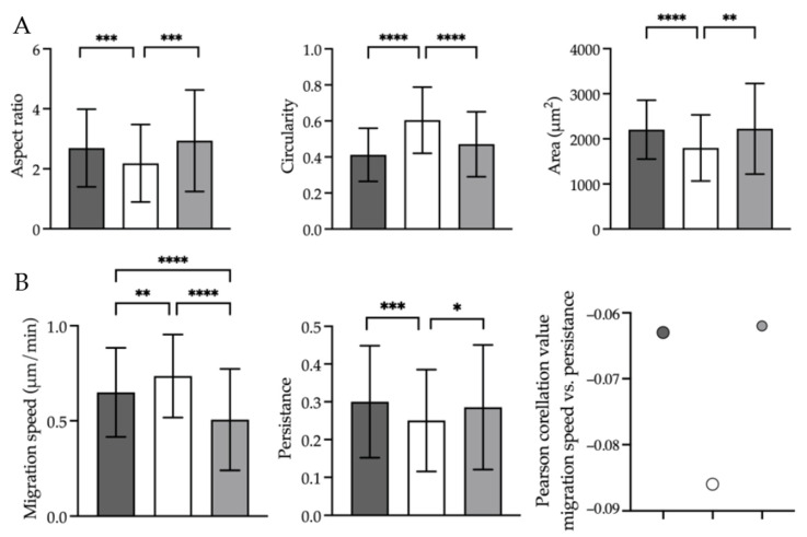
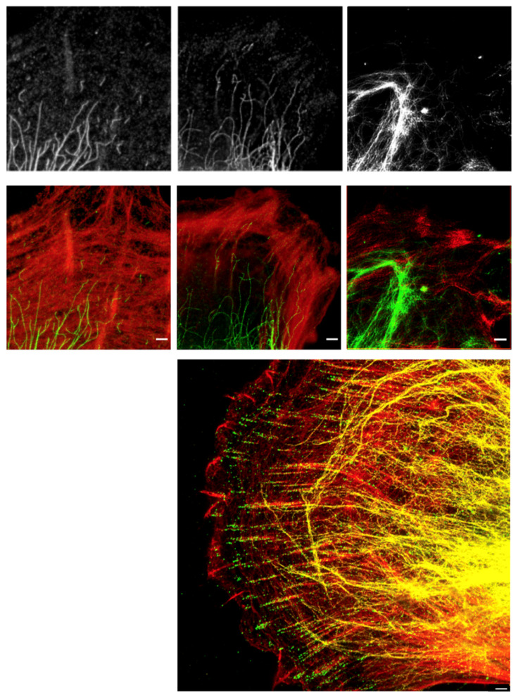
Metastasising cells express the intermediate filament protein vimentin, which is used to diagnose invasive tumours in the clinic. We aimed to clarify how vimentin regulates the motility of metastasising fibroblasts. STED super-resolution microscopy, live-cell imaging and quantitative proteomics revealed that oncogene-expressing and metastasising fibroblasts show a less-elongated cell shape, reduced cell spreading, increased cell migration speed, reduced directionality, and stronger coupling between these migration parameters compared to normal control cells. In total, we identified and compared 555 proteins in the vimentin interactome. In metastasising cells, the levels of keratin 18 and Rab5C were increased, while those of actin and collagen were decreased. Inhibition of HDAC6 reversed the shape, spreading and migration phenotypes of metastasising cells back to normal. Inhibition of HDAC6 also decreased the levels of talin 1, tropomyosin, Rab GDI β, collagen and emilin 1 in the vimentin interactome, and partially reversed the nanoscale vimentin organisation in oncogene-expressing cells. These findings describe the changes in the vimentin interactome and nanoscale distribution that accompany the defective cell shape, spreading and migration of metastasising cells. These results support the hypothesis that oncogenes can act through HDAC6 to regulate the vimentin binding of the cytoskeletal and cell-extracellular matrix adhesion components that contribute to the defective motility of metastasising cells.
转移细胞表达中间丝蛋白波形蛋白,临床上用于诊断侵袭性肿瘤。我们旨在阐明波形蛋白如何调节转移成纤维细胞的运动性。STED 超分辨率显微镜、活细胞成像和定量蛋白质组学揭示,与正常对照细胞相比,表达致癌基因和转移的成纤维细胞表现出更短的细胞形状、减少的细胞铺展、增加的细胞迁移速度、降低的方向性以及这些迁移参数之间更强的耦合。总共,我们在波形蛋白互作组中鉴定和比较了 555 种蛋白质。在转移细胞中,角蛋白 18 和 Rab5C 的水平增加,而肌动蛋白和胶原蛋白的水平降低。抑制 HDAC6 将转移细胞的形状、铺展和迁移表型逆转回正常。抑制 HDAC6 还降低了波形蛋白互作组中 talin 1、原肌球蛋白、Rab GDI β、胶原蛋白和 emilin 1 的水平,并部分逆转了表达致癌基因的细胞中纳米尺度波形蛋白的组织。这些发现描述了伴随转移细胞缺陷形状、铺展和迁移的波形蛋白互作组和纳米尺度分布的变化。这些结果支持这样一种假设,即致癌基因可以通过 HDAC6 来调节细胞骨架和细胞-细胞外基质黏附成分与波形蛋白的结合,从而导致转移细胞运动性缺陷。