Gynecologic Oncology Laboratory, Brigham and Women's Hospital, 75 Francis Street, Boston, MA, 02132, USA.
PNP Research Corporation, LLC, 282 S COUNTY RD, Drury, MA, 01343, USA.
J Ovarian Res. 2022 Feb 26;15(1):28. doi: 10.1186/s13048-022-00957-7.
Measurement of serum CA125, an antigenic fragment of human mucin 16 (MUC16), is used to monitor the clinical progression of epithelial ovarian cancer (EOC). However, rather than simply a passive marker reflecting tumor burden, MUC16 may have a more active role by binding to immune cells and altering their tumor response. We developed a research tool to measure MUC16-binding to the surfaces of peripheral blood mononuclear cell (PBMC) subtypes and tested its research value using specimens collected serially from a woman being treated for high grade serous EOC.
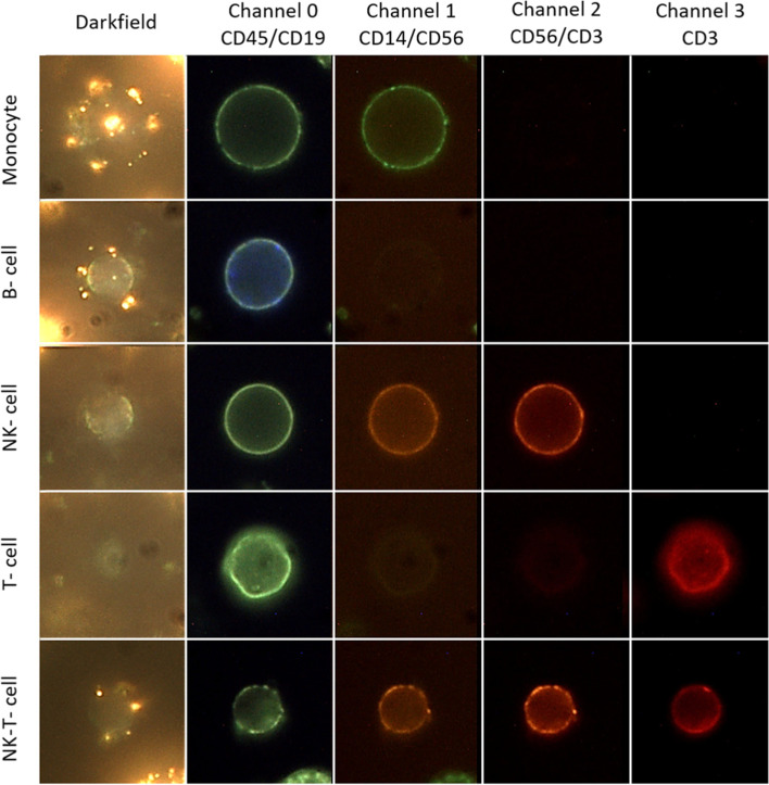
Cryopreserved PBMCs were mixed with anti-CA125 antibody-labeled plasmonic gold nanoparticles (PNPs) to detect cell surface MUC16-binding along with fluorescent stains to identify B cells, NK cells, NK-T cells, T cells, and monocytes. From 3D darkfield images, a computer algorithm was applied to enumerate PNP-binding and fluorescence microscopy to identify cell lineage. Average MUC16-binding was determined by fitting a Poisson distribution to PNP-counts across similar cell types. MUC16-binding to cell types was correlated with treatment details, CA125 levels, and complete blood count (CBC) data.
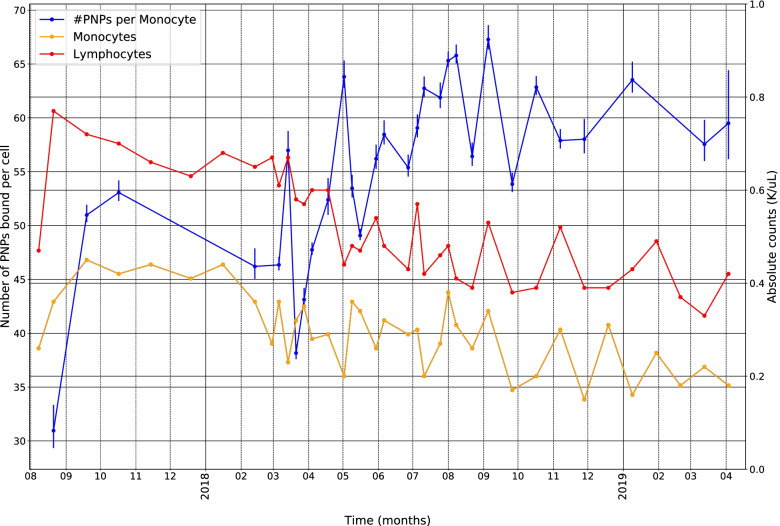
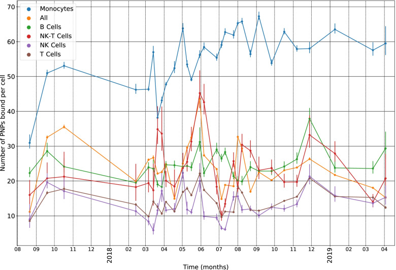
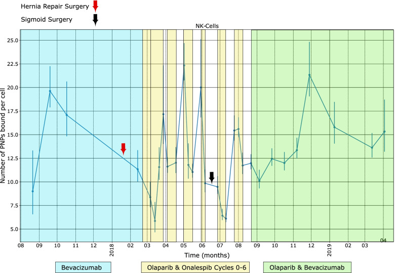
Over a 21-month period, monocytes had the highest level of MUC16-binding which was positively correlated with serum CA125 and inversely correlated with circulating monocyte and lymphocyte counts. Fluctuations of PNP-binding to NK cells were associated temporally with types of chemotherapy and surgical events. Levels of MUC16 bound to NK cells were positively correlated with levels of MUC16 bound to T and NK-T cells and inversely correlated with circulating platelets.
Assessment of MUC16-binding among cryopreserved PBMC cell types can be accomplished using darkfield and fluorescence microscopy. Correlations observed between level of binding by cell type with serum CA125, CBC data, and treatment details suggest that the new techniques may offer novel insights into EOC's clinical course.
血清 CA125 的测量,作为人类黏蛋白 16(MUC16)的抗原片段,用于监测上皮性卵巢癌(EOC)的临床进展。然而,MUC16 可能具有更积极的作用,它不仅简单地作为反映肿瘤负荷的被动标志物,还可以与免疫细胞结合并改变其对肿瘤的反应。我们开发了一种研究工具来测量外周血单个核细胞(PBMC)亚群表面上的 MUC16 结合,并使用从一名正在接受高级别浆液性 EOC 治疗的女性中连续采集的标本测试其研究价值。
将冷冻保存的 PBMC 与抗 CA125 抗体标记的等离子体金纳米颗粒(PNP)混合,以检测细胞表面 MUC16 结合,同时用荧光染料鉴定 B 细胞、NK 细胞、NK-T 细胞、T 细胞和单核细胞。从 3D 暗场图像中,应用计算机算法对 PNP 结合进行计数,并通过荧光显微镜识别细胞谱系。通过对类似细胞类型的 PNP 计数进行泊松分布拟合,确定 MUC16 结合的平均值。将 MUC16 与细胞类型的结合与治疗细节、CA125 水平和全血细胞计数(CBC)数据相关联。
在 21 个月的时间里,单核细胞具有最高水平的 MUC16 结合,与血清 CA125 呈正相关,与循环单核细胞和淋巴细胞计数呈负相关。PNP 与 NK 细胞结合的波动与化疗和手术事件的类型具有时间相关性。NK 细胞结合的 MUC16 水平与 T 和 NK-T 细胞结合的 MUC16 水平呈正相关,与循环血小板呈负相关。
使用暗场和荧光显微镜可以完成对冷冻保存的 PBMC 细胞类型中 MUC16 结合的评估。观察到的与细胞类型结合水平与血清 CA125、CBC 数据和治疗细节之间的相关性表明,新技术可能为 EOC 的临床过程提供新的见解。