Guo Yuanhao, Shen Di, Zhou Yanfeng, Yang Yutong, Liang Jinzhao, Zhou Yating, Li Ningning, Liu Yu, Yang Ge, Li Wenjing
Laboratory of Computational Biology and Machine Intelligence, National Laboratory of Pattern Recognition, Institute of Automation, Chinese Academy of Sciences, Beijing, China.
School of Artificial Intelligence, University of Chinese Academy of Sciences, Beijing, China.
Front Cell Dev Biol. 2022 Jan 21;9:767866. doi: 10.3389/fcell.2021.767866. eCollection 2021.
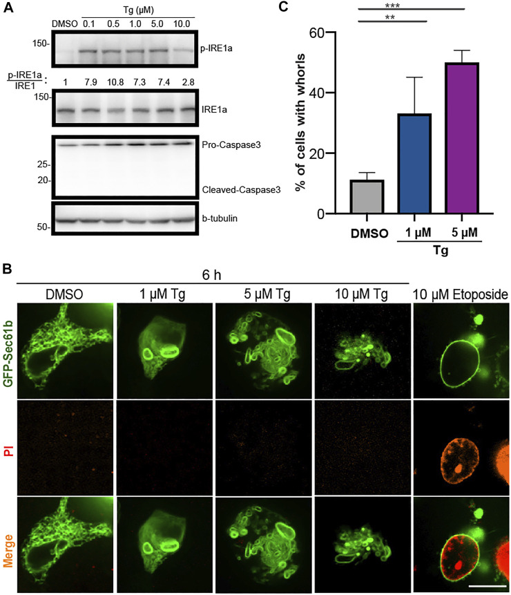
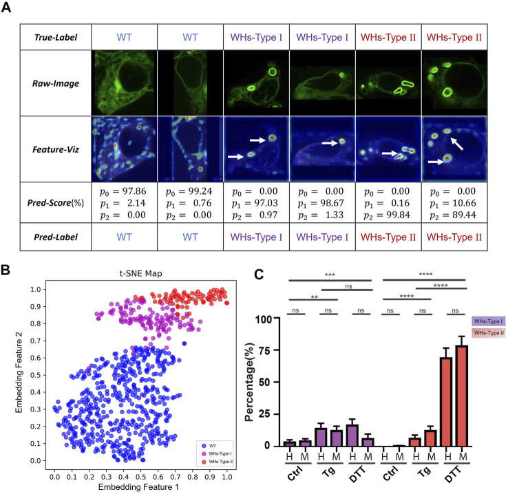
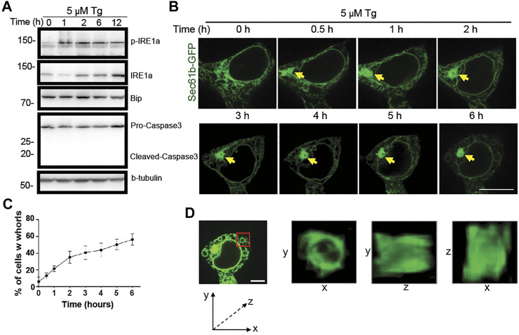
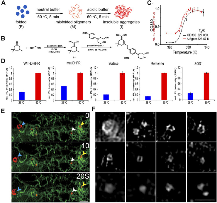
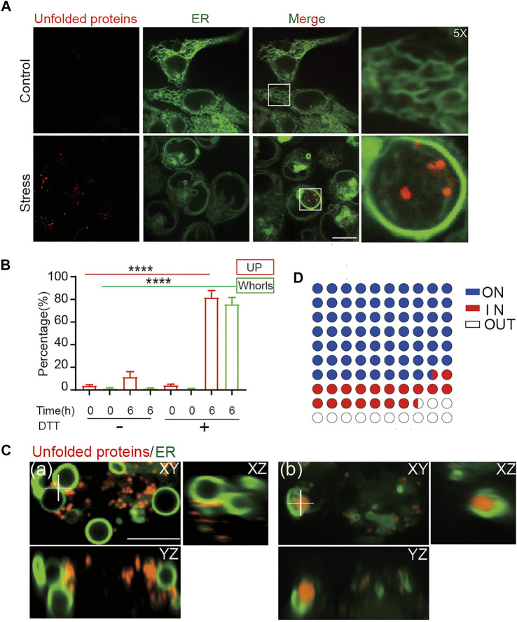
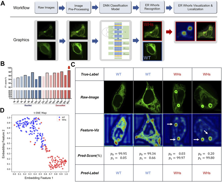
Endoplasmic reticulum stress (ER stress) is a condition that is defined by abnormal accumulation of unfolded proteins. It plays an important role in maintaining cellular protein, lipid, and ion homeostasis. By triggering the unfolded protein response (UPR) under ER stress, cells restore homeostasis or undergo apoptosis. Chronic ER stress is implicated in many human diseases. Despite extensive studies on related signaling mechanisms, reliable image biomarkers for ER stress remain lacking. To address this deficiency, we have validated a morphological image biomarker for ER stress and have developed a deep learning-based assay to enable automated detection and analysis of this marker for screening studies. Specifically, ER under stress exhibits abnormal morphological patterns that feature ring-shaped structures called whorls (WHs). Using a highly specific chemical probe for unfolded and aggregated proteins, we find that formation of ER whorls is specifically associated with the accumulation of the unfolded and aggregated proteins. This confirms that ER whorls can be used as an image biomarker for ER stress. To this end, we have developed ER-WHs-Analyzer, a deep learning-based image analysis assay that automatically recognizes and localizes ER whorls similarly as human experts. It does not require laborious manual annotation of ER whorls for training of deep learning models. Importantly, it reliably classifies different patterns of ER whorls induced by different ER stress drugs. Overall, our study provides mechanistic insights into morphological patterns of ER under stress as well as an image biomarker assay for screening studies to dissect related disease mechanisms and to accelerate related drug discoveries. It demonstrates the effectiveness of deep learning in recognizing and understanding complex morphological phenotypes of ER.
内质网应激(ER应激)是一种由未折叠蛋白异常积累所定义的状态。它在维持细胞蛋白质、脂质和离子稳态中发挥重要作用。通过在ER应激下触发未折叠蛋白反应(UPR),细胞可恢复稳态或发生凋亡。慢性ER应激与许多人类疾病有关。尽管对相关信号机制进行了广泛研究,但仍缺乏可靠的ER应激图像生物标志物。为解决这一缺陷,我们验证了一种用于ER应激的形态学图像生物标志物,并开发了一种基于深度学习的检测方法,以实现对该标志物的自动检测和分析,用于筛选研究。具体而言,应激状态下的内质网呈现出异常的形态模式,其特征是具有称为涡旋(WHs)的环形结构。使用针对未折叠和聚集蛋白的高度特异性化学探针,我们发现内质网涡旋的形成与未折叠和聚集蛋白的积累特异性相关。这证实了内质网涡旋可作为ER应激的图像生物标志物。为此,我们开发了ER-WHs-Analyzer,这是一种基于深度学习的图像分析检测方法,它能像人类专家一样自动识别和定位内质网涡旋。它不需要为训练深度学习模型而对内质网涡旋进行费力的手动注释。重要的是,它能可靠地对由不同ER应激药物诱导的内质网涡旋的不同模式进行分类。总体而言,我们的研究为应激状态下内质网的形态模式提供了机制性见解,以及一种用于筛选研究的图像生物标志物检测方法,以剖析相关疾病机制并加速相关药物发现。它证明了深度学习在识别和理解内质网复杂形态表型方面的有效性。